Biopolis Dresden Imaging Platform

Gallery

Here all images used on this website can be found including the author, the title and a short description. If you want to know more details about any of those, just get in touch with us!

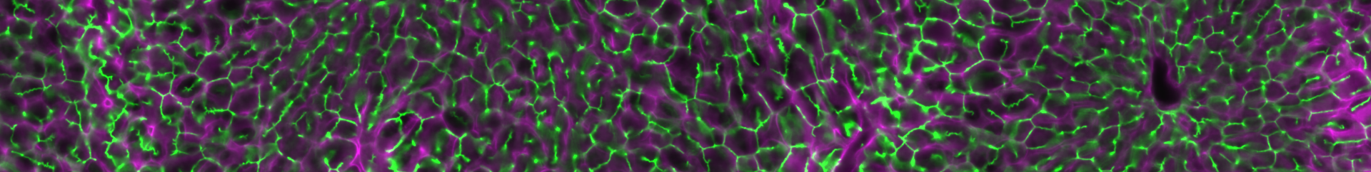
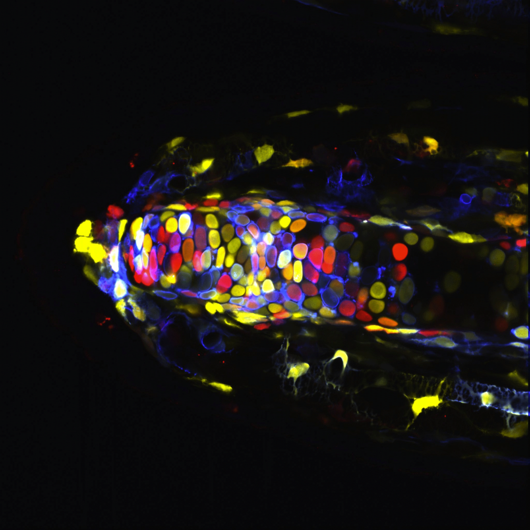
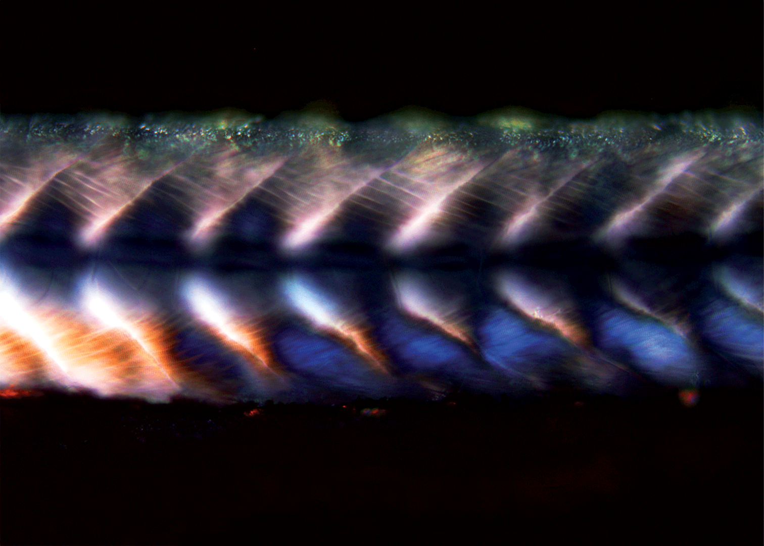
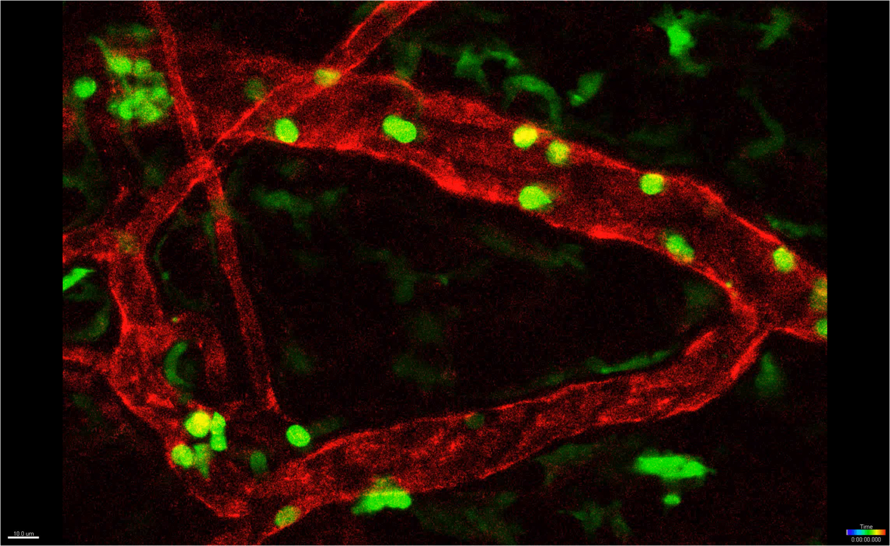
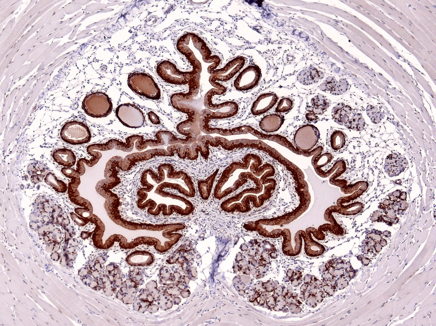
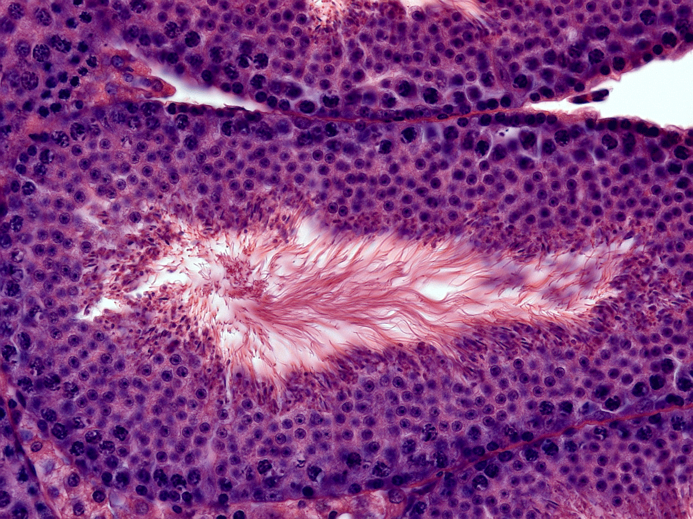
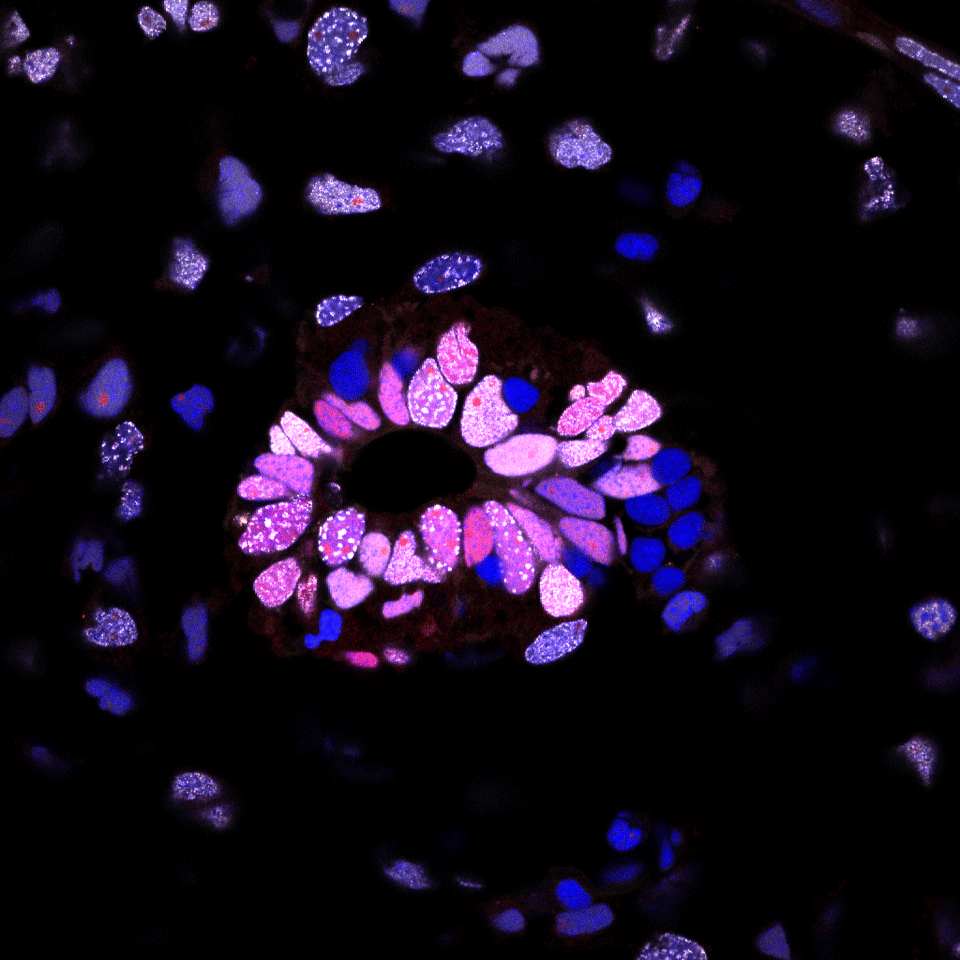
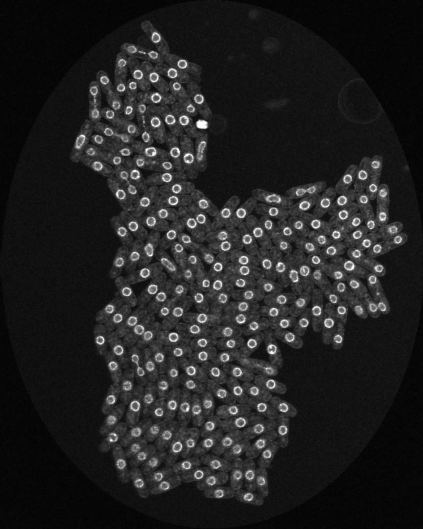
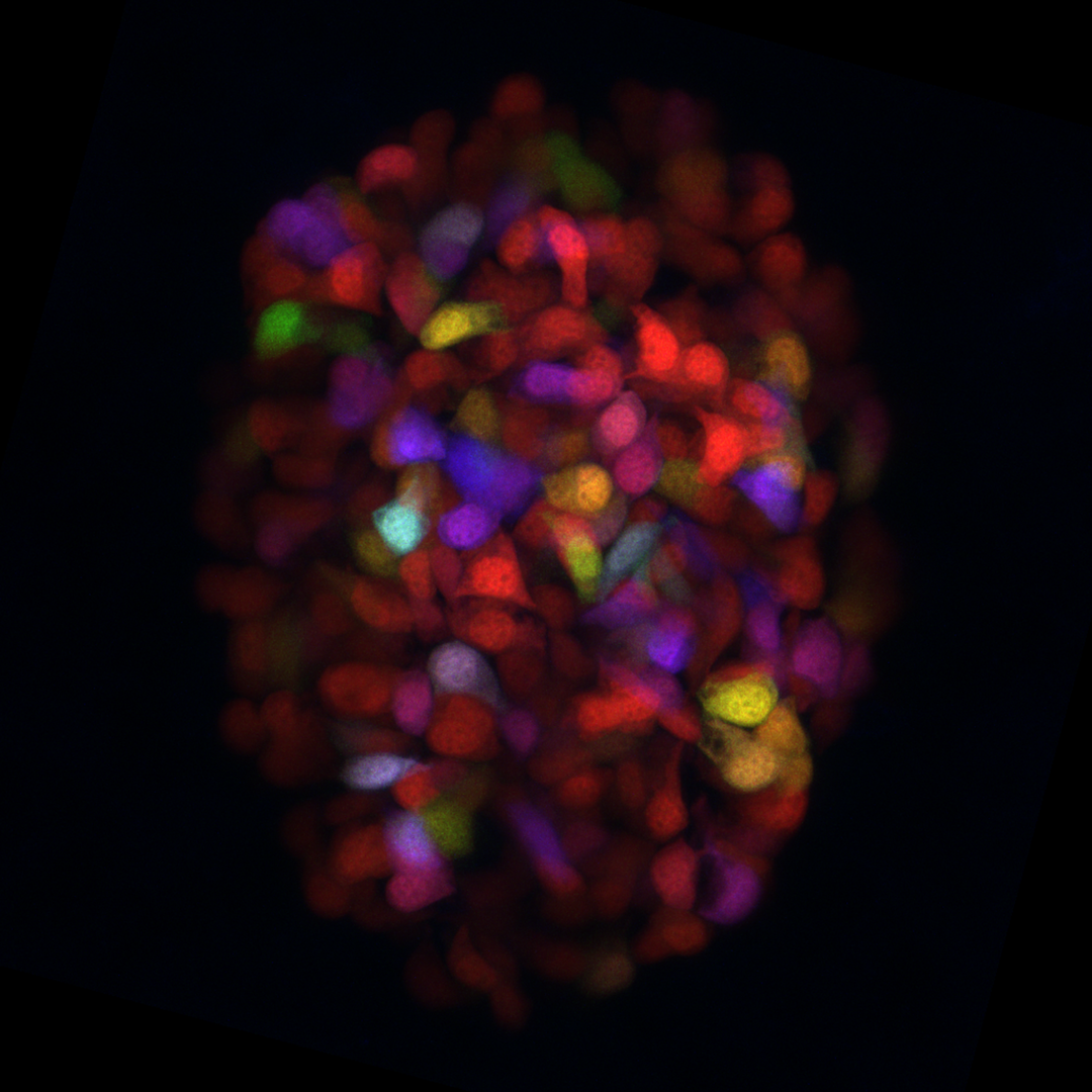
Light Microscopy Images

Josh Currie - CTRD - Axolotl tissue expressing a multicolor fluorescent LoxP reporter (i.e. Brainbow technology) supported by LMF CRTD/BIOTEC
Axolotl tissue expressing a multicolor fluorescent LoxP reporter (i.e. Brainbow technology) supported by LMF CRTD/BIOTEC
Judith Konantz - CRTD - sceleton muscles in a zebrafish larva imaged with polarized light microscopy, supported by LMF CRTD/BIOTEC
A 5-day-old zebrafish larva visualized by polarized light microscopy. The birefrigence of the muscle enables to observe the living larva without any staining, supported by LMF CRTD/BIOTEC
Jan Dudeck - MTZ Immunology - Mouse skin blood vessel
The circulation of neutrophil granulocytes in a mouse skin blood vessel. The image has been acquired in a living mouse, supported by MTZ, Z3-Facility
Edgar Büttner - BIOTEC - Utethra
The image shows a urethra acquired with brightfield microscopy, supported by LMF CRTD/BIOTEC
Edgar Büttner - BIOTEC - Testis
The image shows histological stained testis acquired with brightfield microscopy, supported by LMF CRTD/BIOTEC
© Jan Dudeck - MTZ,  Immunology - Mouse lymph node
A multi colour image of a mouse inguinal lymph node. The image has been acquired with mulit photon microscopy in a living mouse, supported by MTZ, Z3-Facility
Kathleen Roensch - CRTD - Spinal cord
Cross section of a regenerating spinal cord. Red: SOX2 (neural stem cell marker); Whit: PCNA (cell proliferation marker); Blue, Hoechst (DNA) supported by LMF CRTD/BIOTEC
Pancreatic beta-cells in zebrafish a colored with rainbow palette.

Electron Microscopy Images
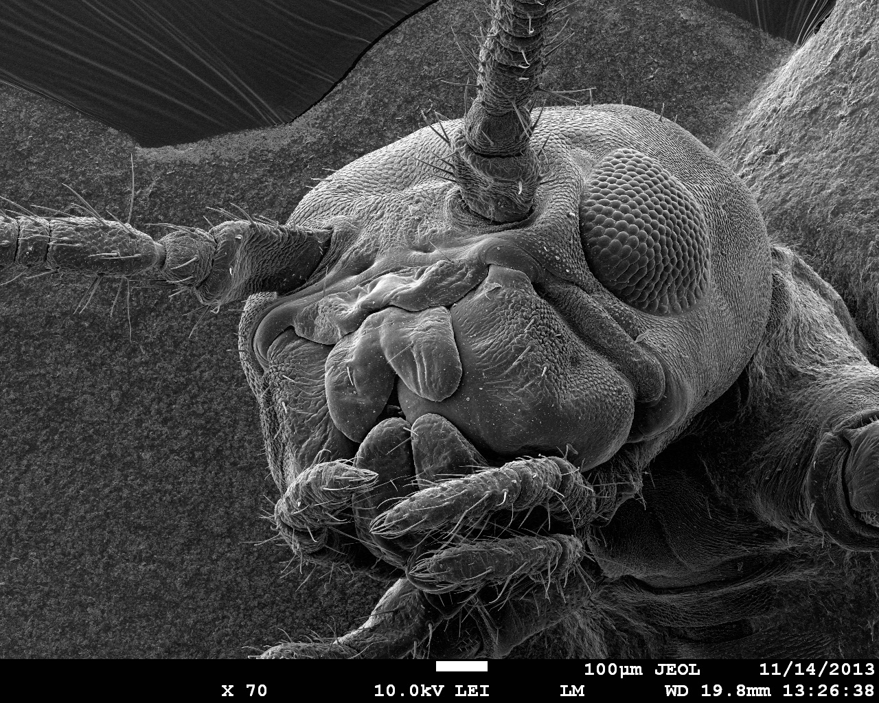
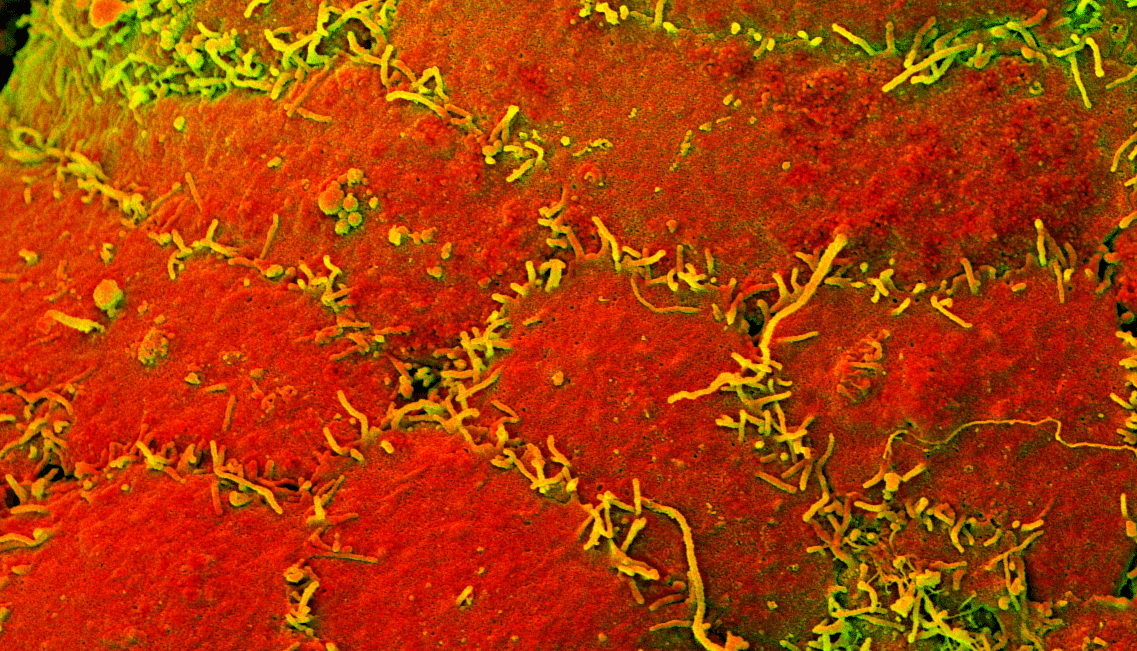
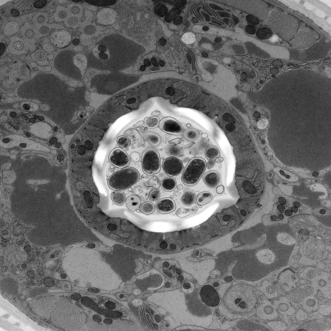
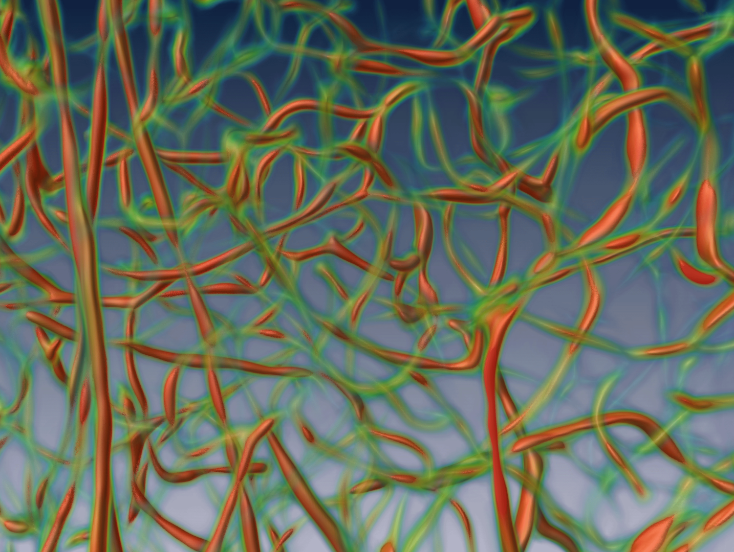
Thomas Kurt - EMF CRTD/BIOTEC - SEM image
SEM image by Thomas Kurth - CRTD
Gokul Kesavan / Michael Brand - CTRD - Brain of a zebrafish embryo
Dorsal view of the brain of a zebrafish embryo. This transgenic embryo expresses a fluorescent protein in its midbrain and in parts of the hindbrain, supported by LMF CRTD/BIOTEC
Thomas Müller-Reichert - MTZ, CFCI  - C.Elegans male
A C.Elegans TEM image by CFCI
Thomas Kurth - EMF CRTD/BIOTEC - Phasmid SEM image
A Phasmid SEM image by EMF CRTD/BIOTEC.
Thomas Kurt - EMF CRTD/BIOTEC - SEM image
SEM image by Thomas Kurth - CRTD

Image Processing

Steffen Vogler / Fanny Boehme - DZNE - Vascular network
3D Reconstruction of vascular network of the dentate gyrus in an adult mouse.

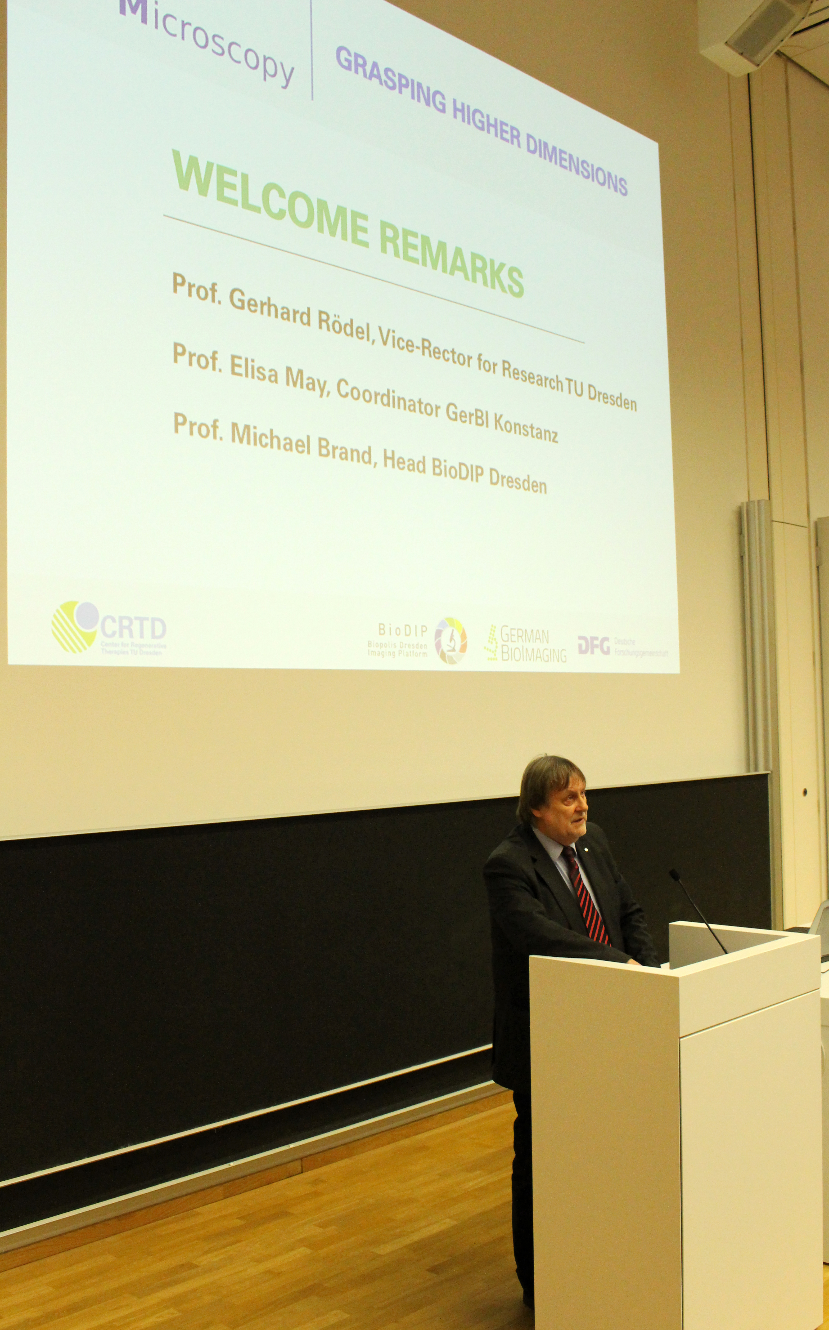
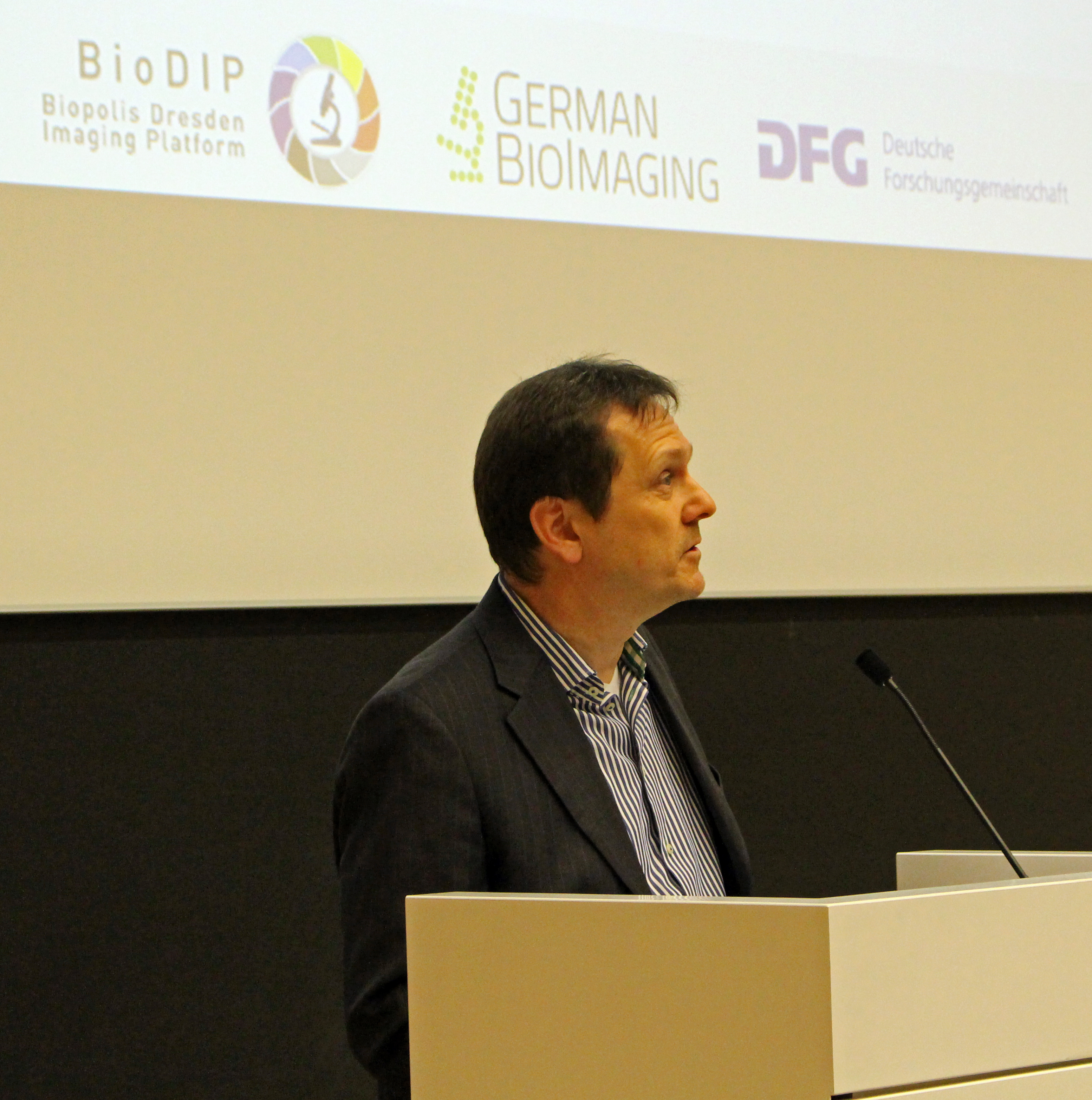
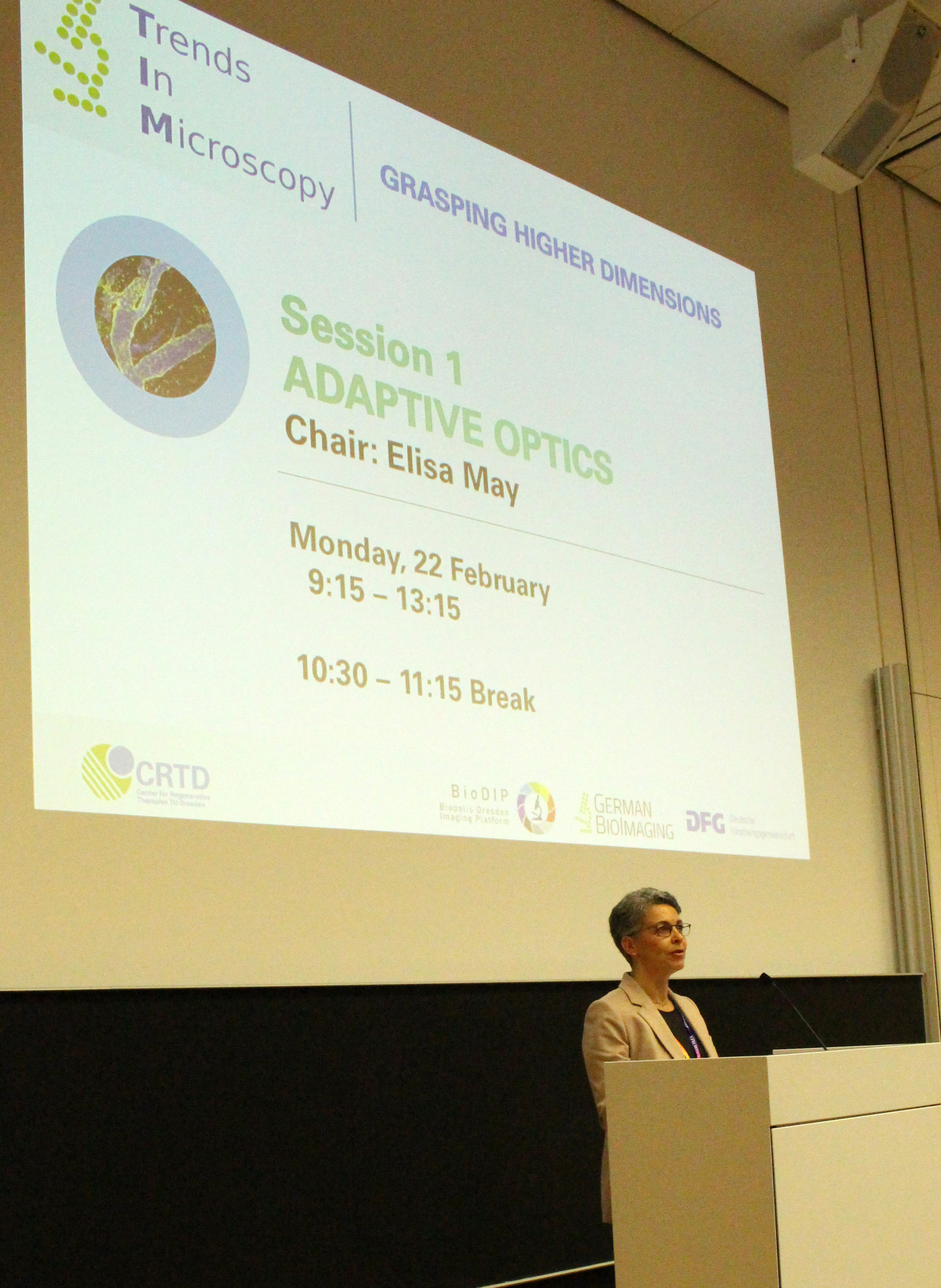
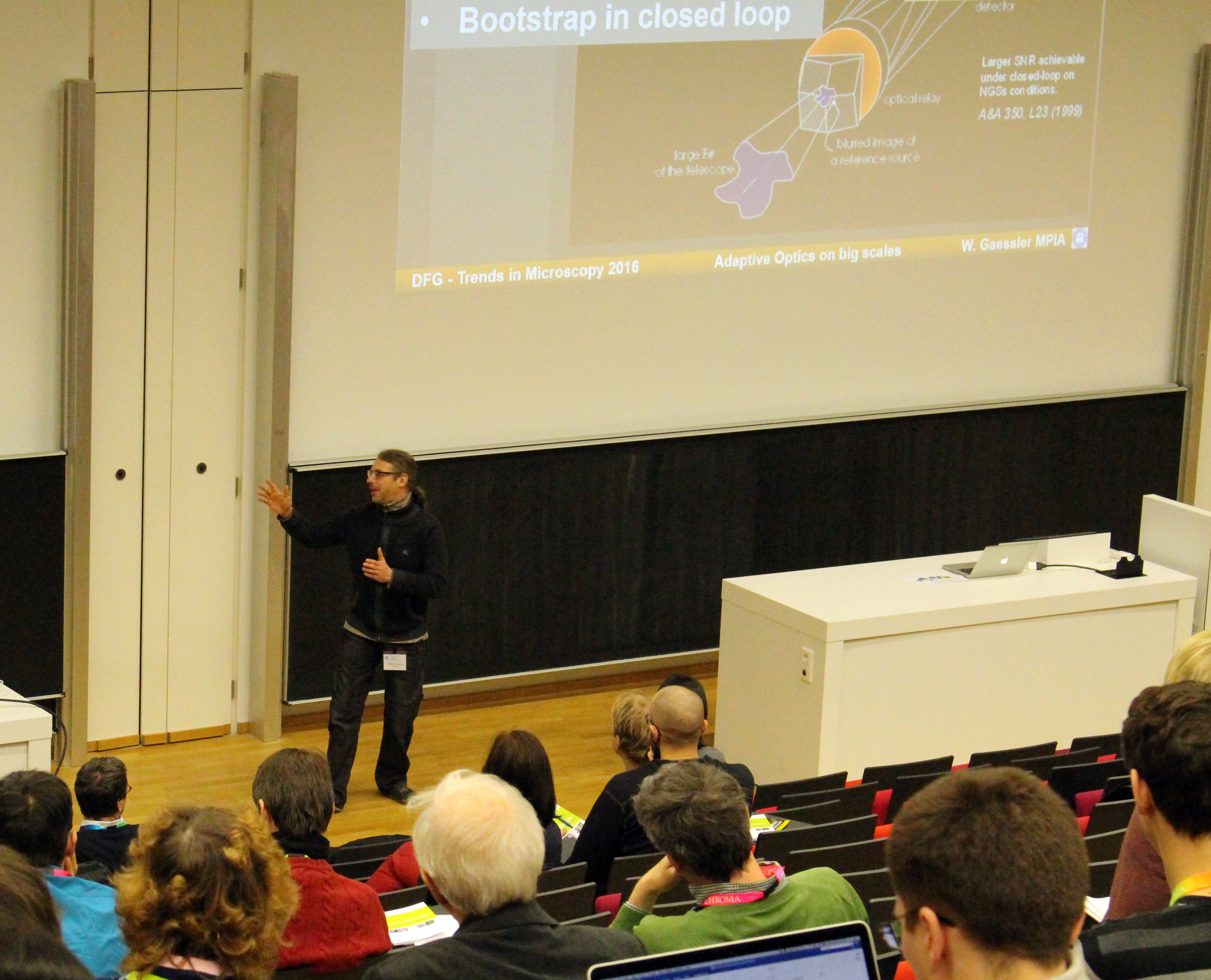
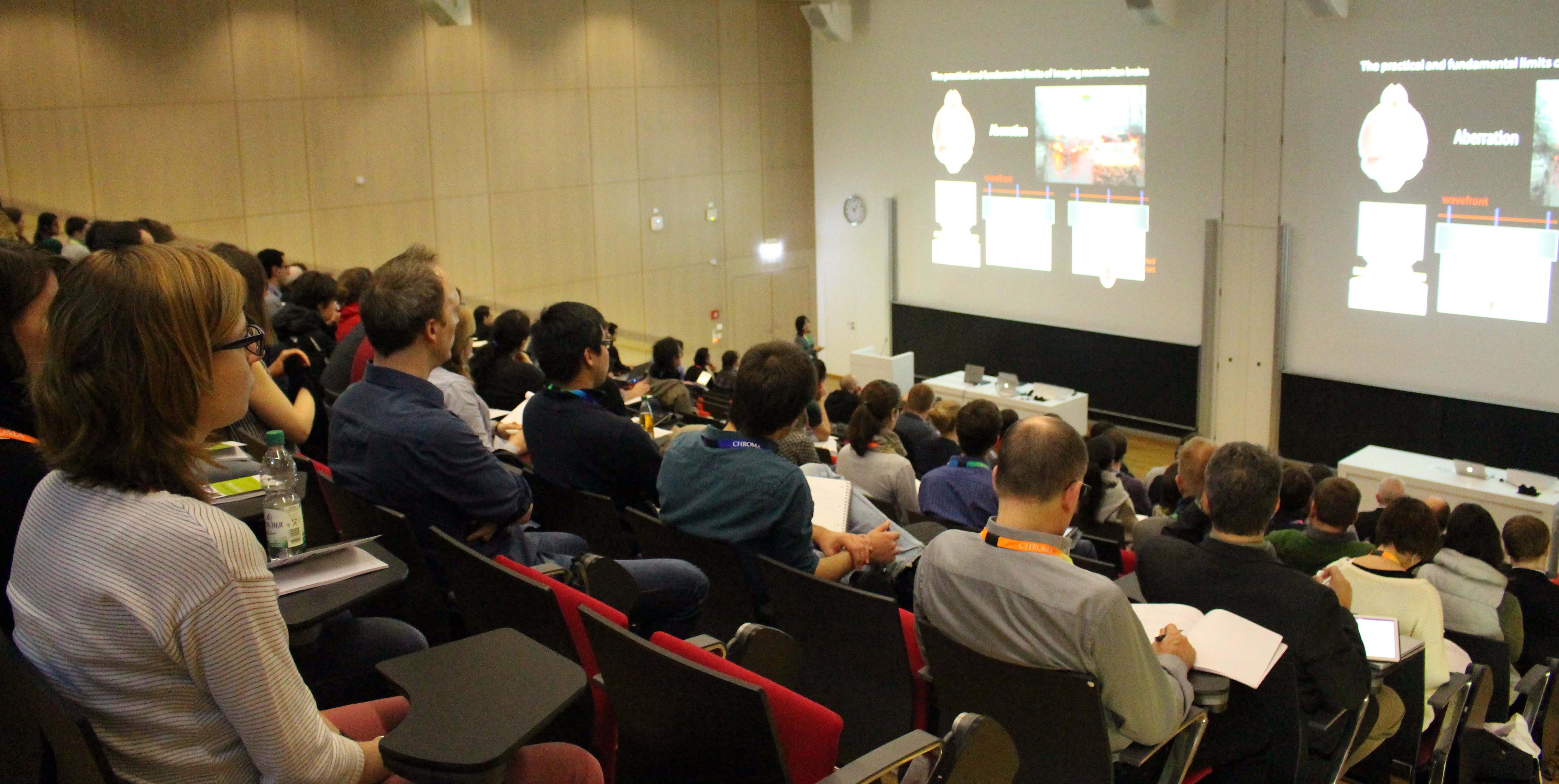
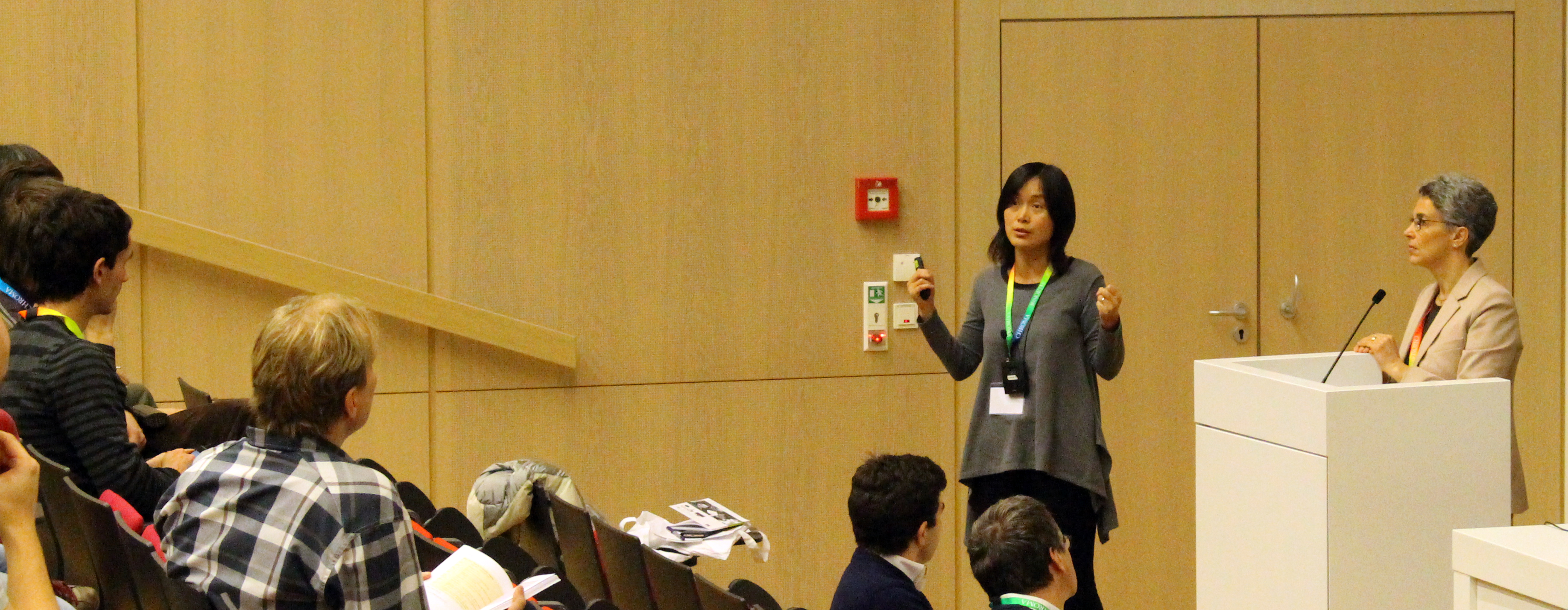
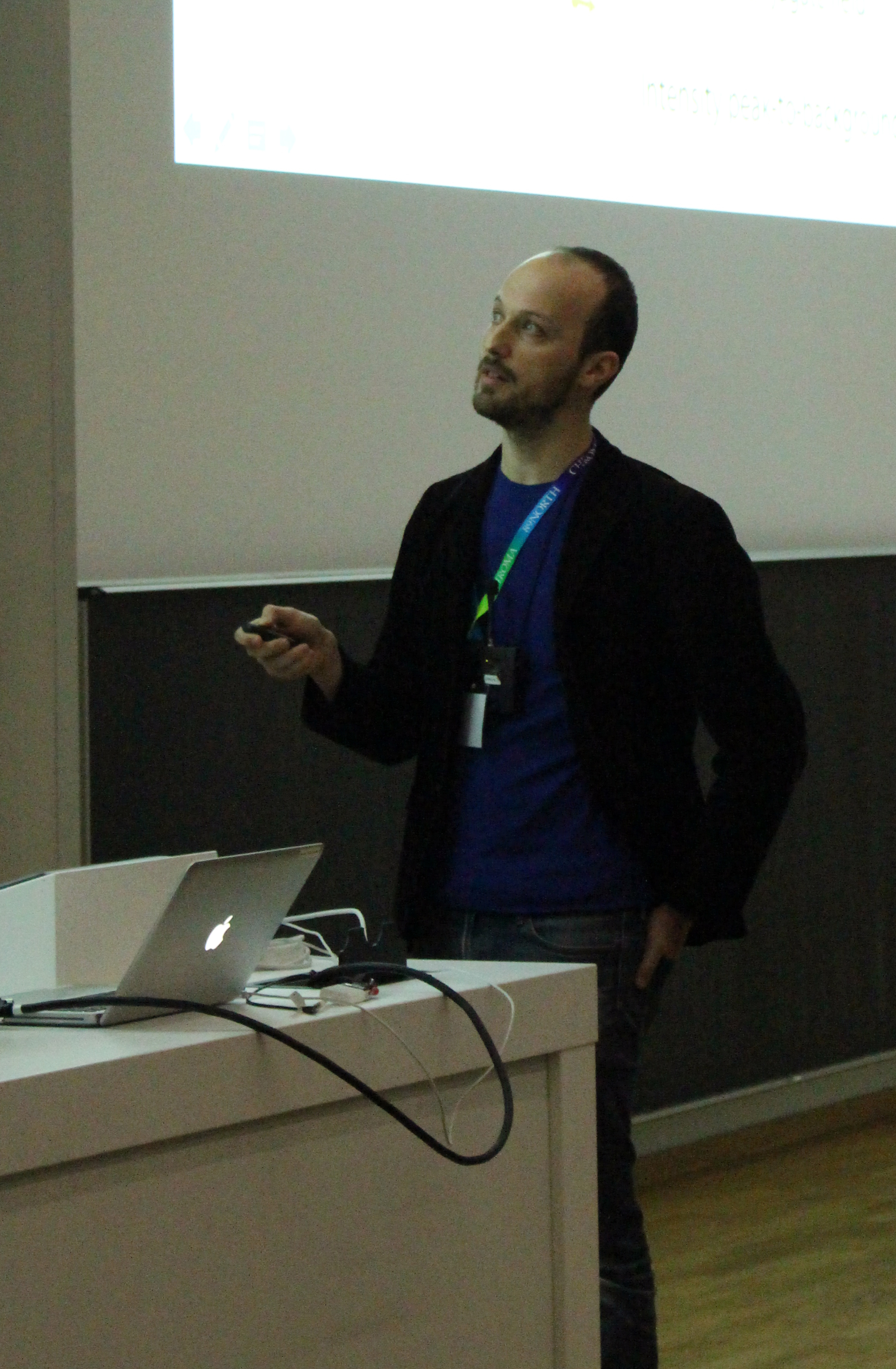
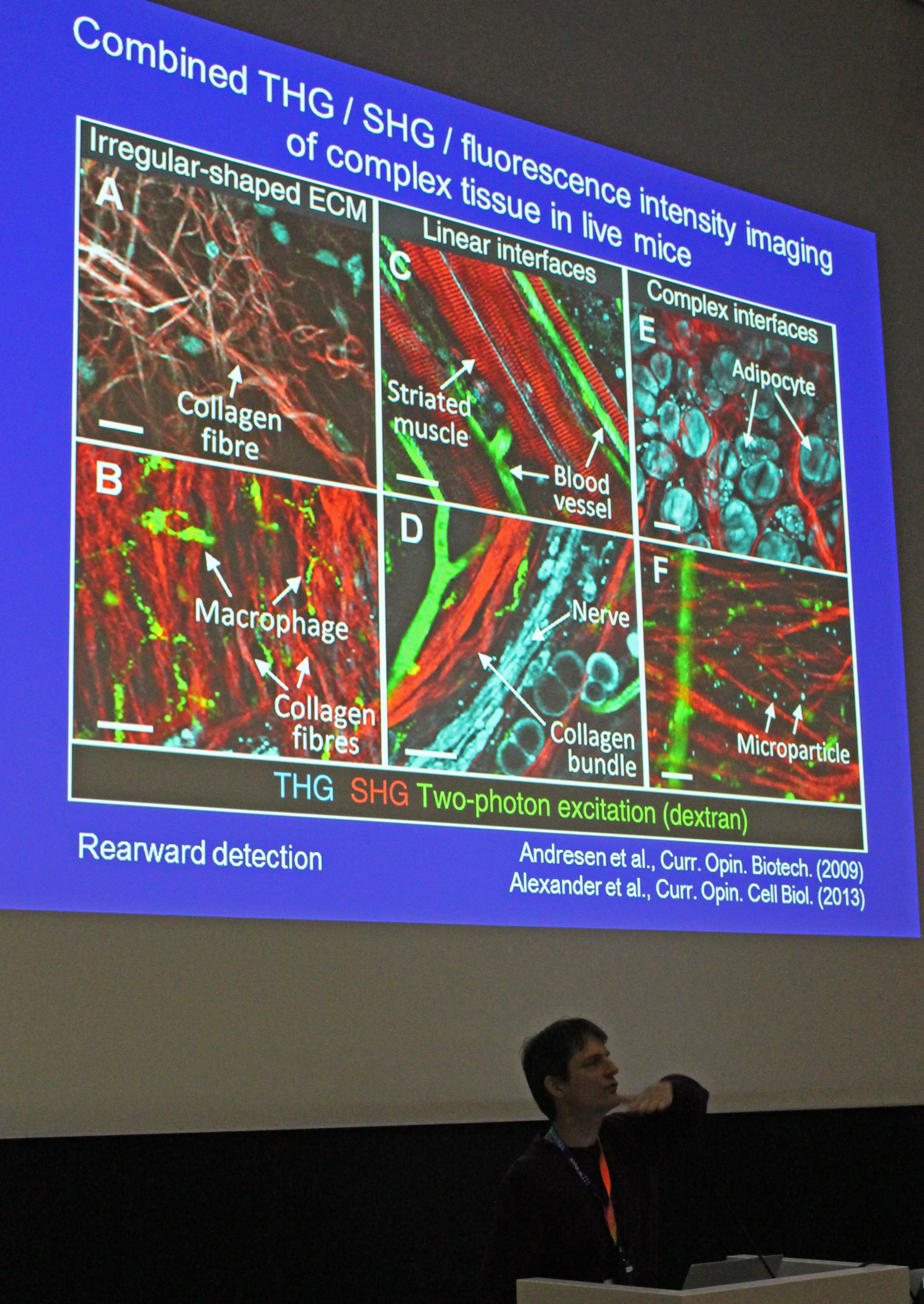
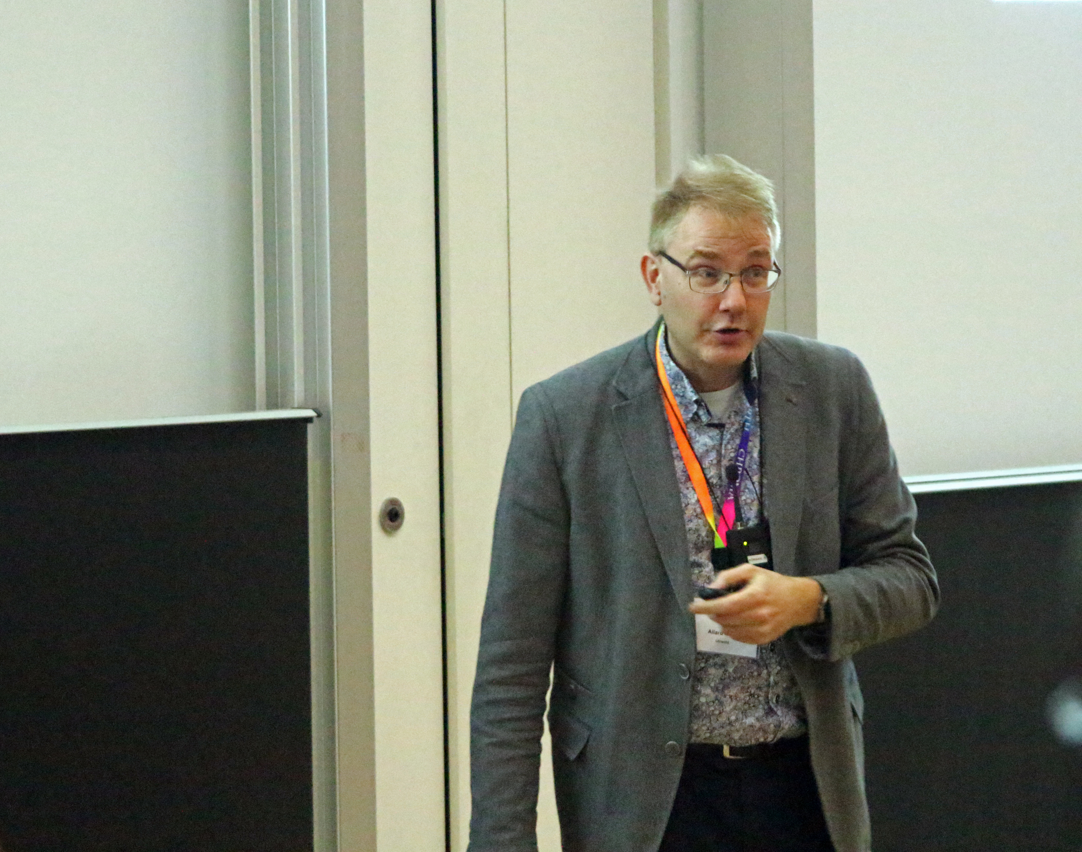
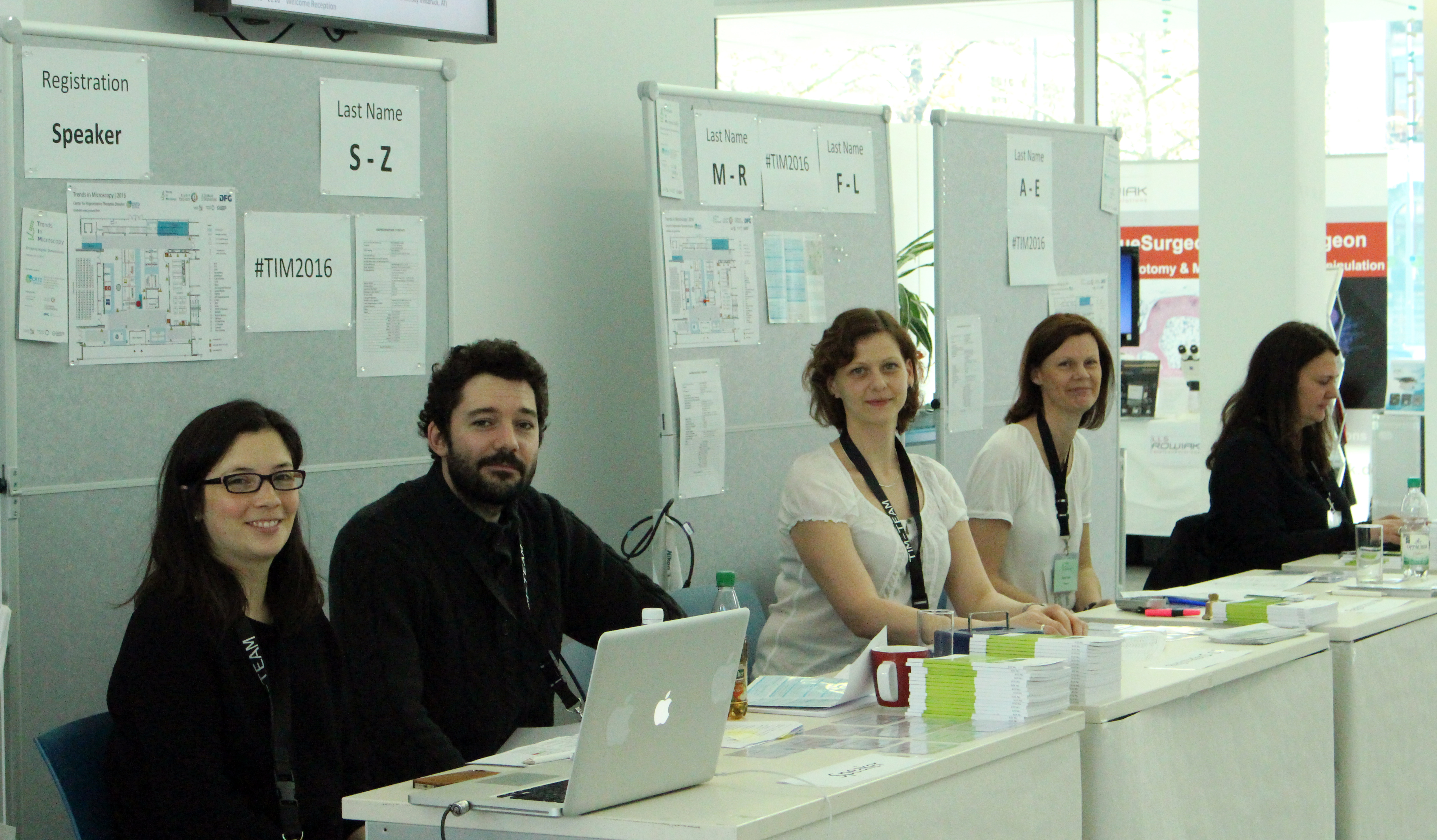
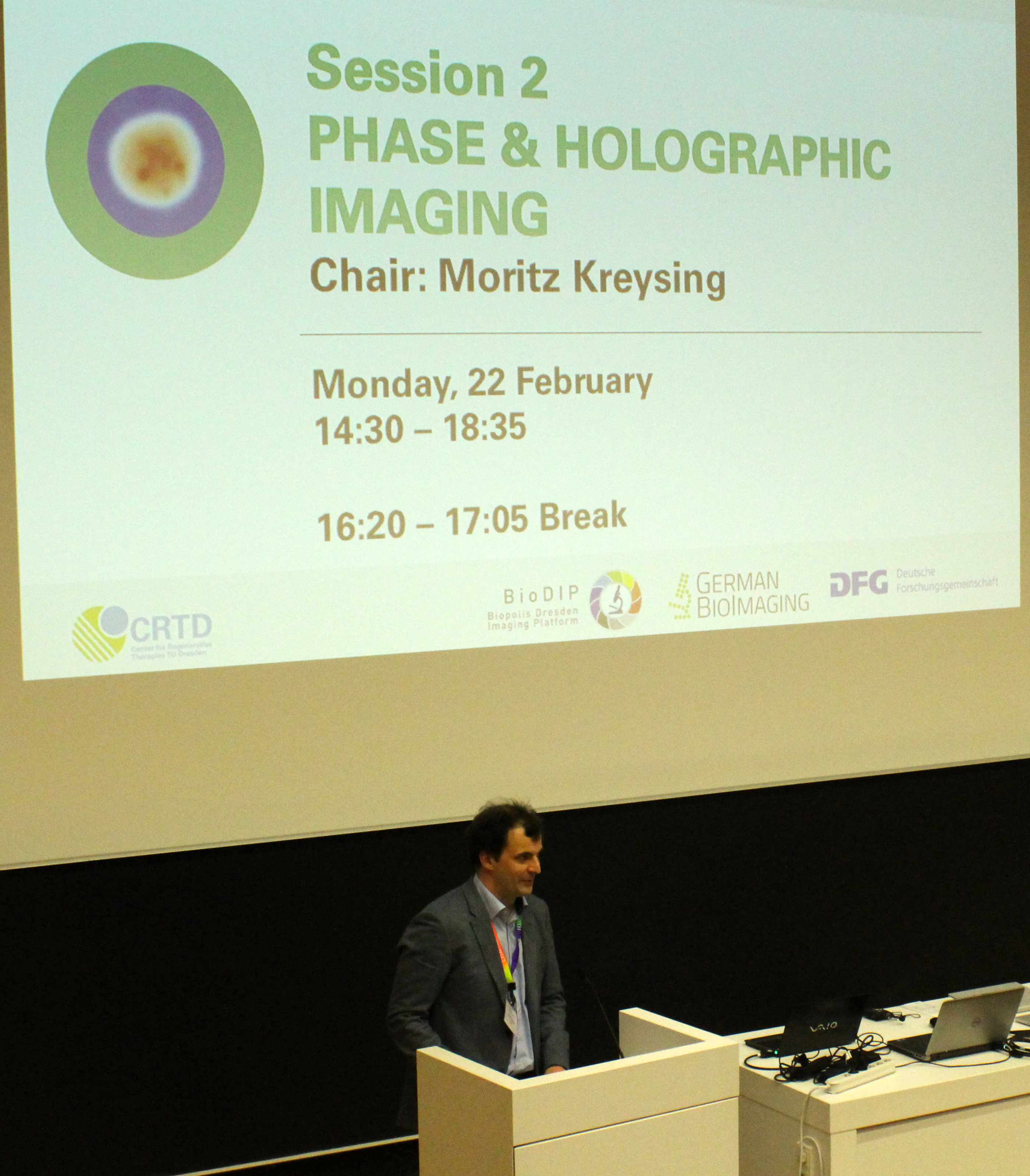
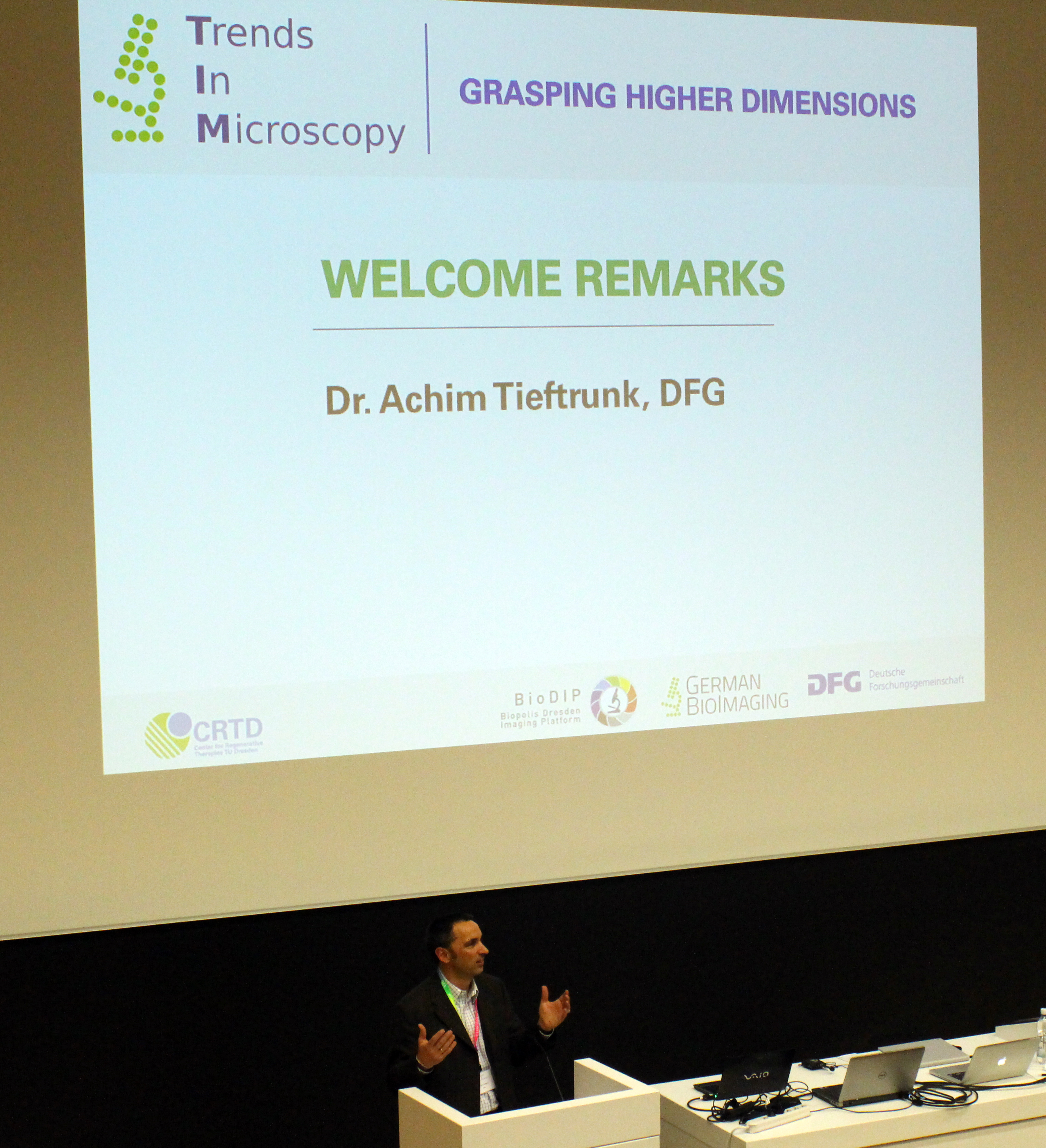
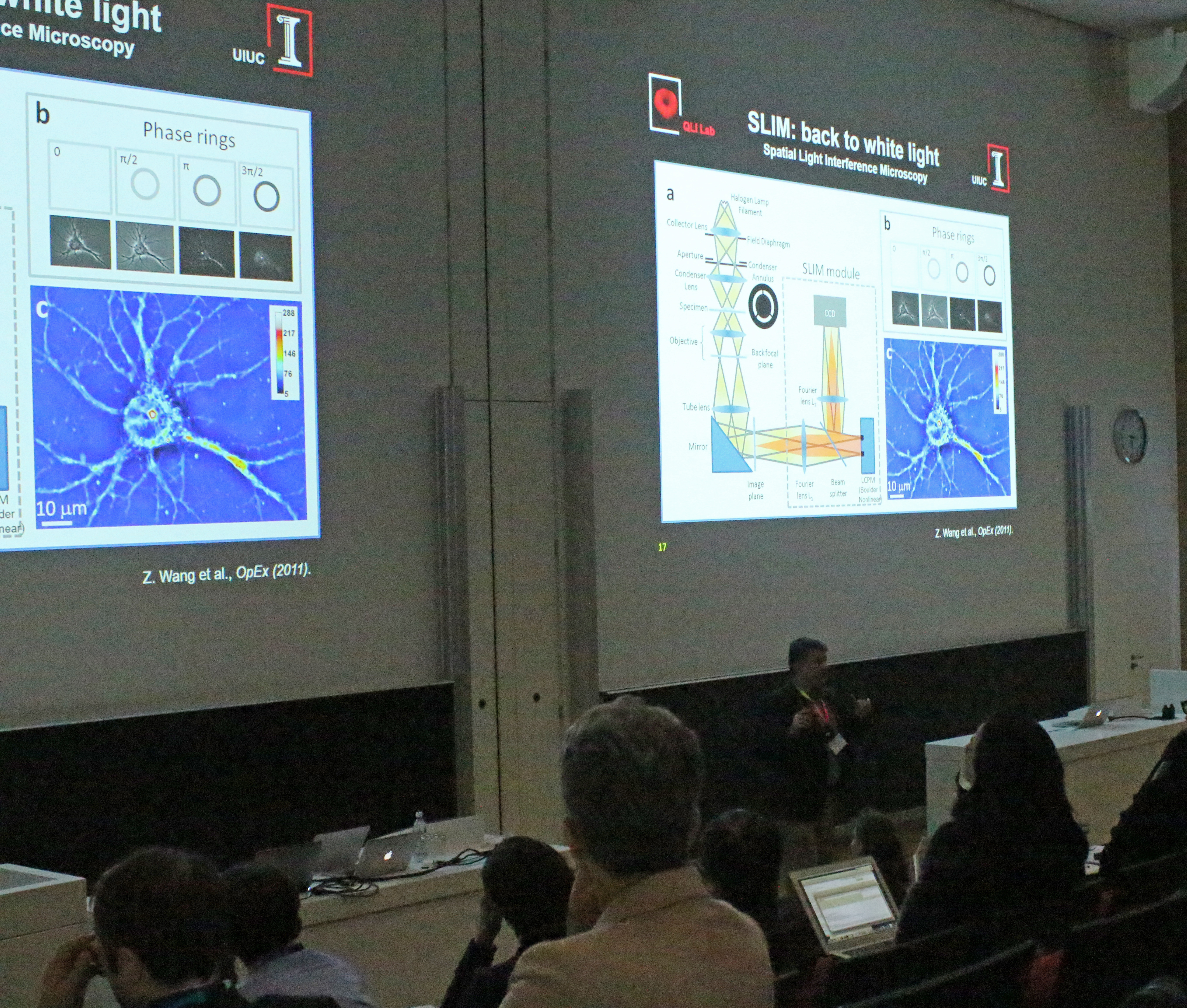
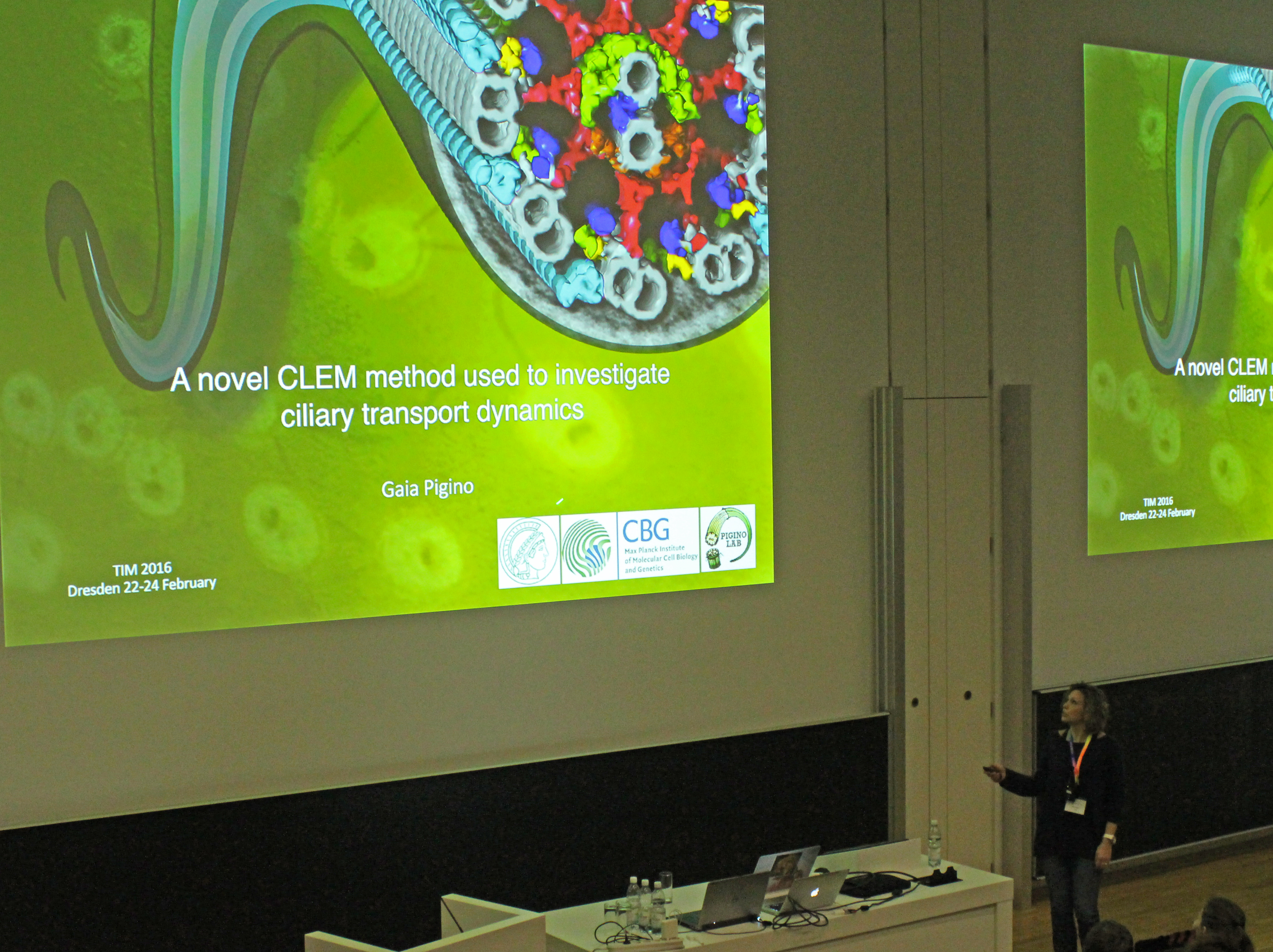
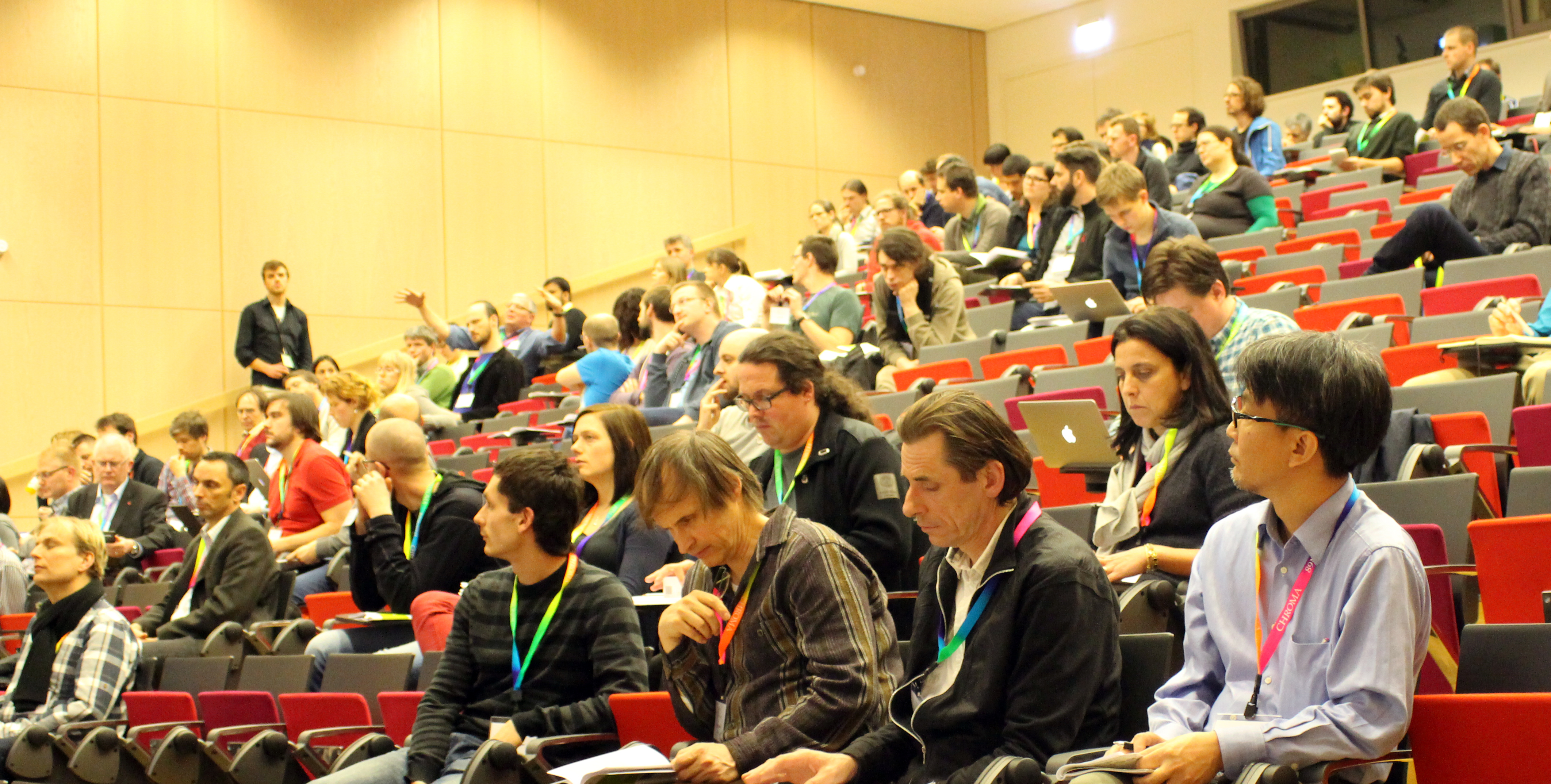
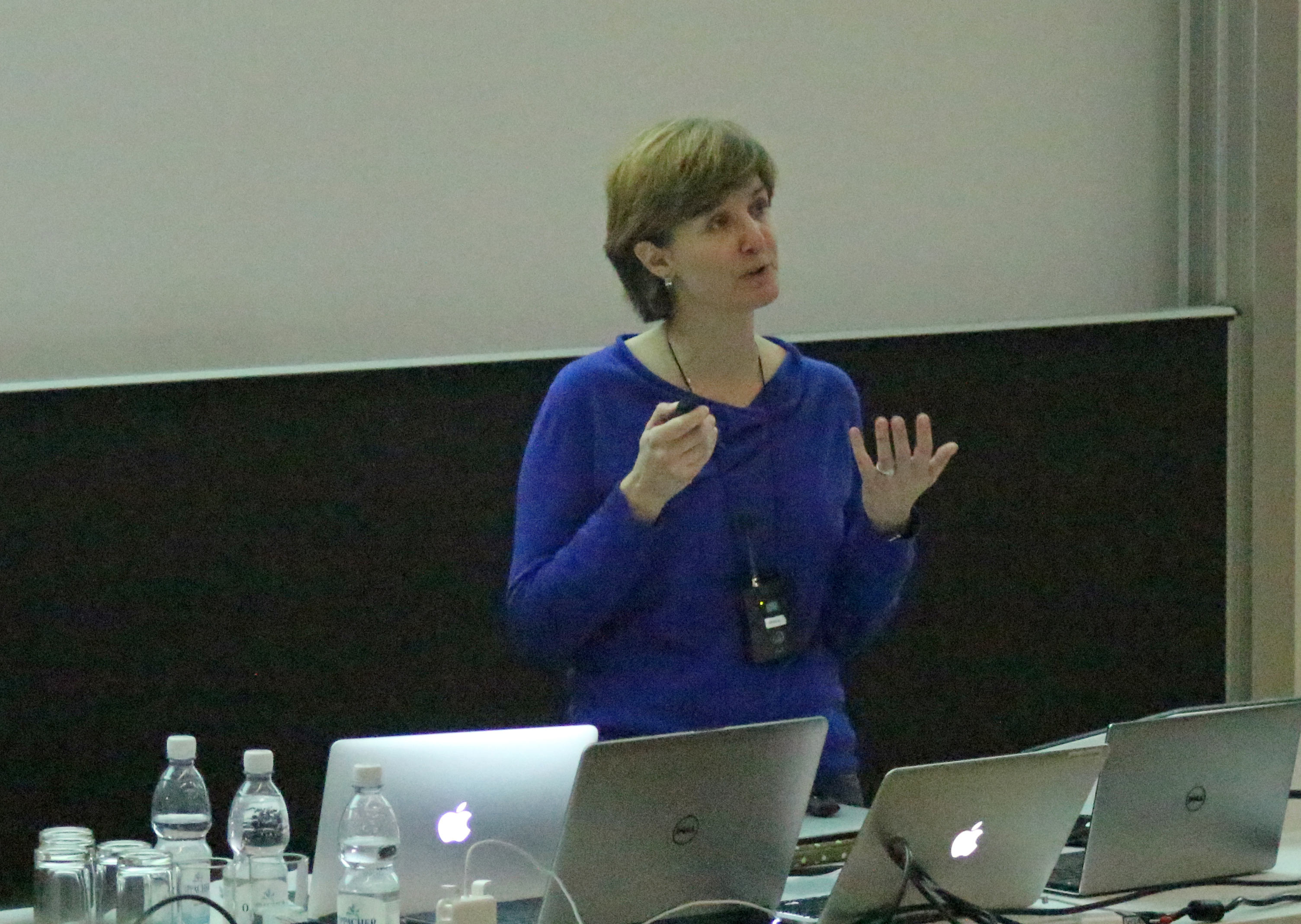
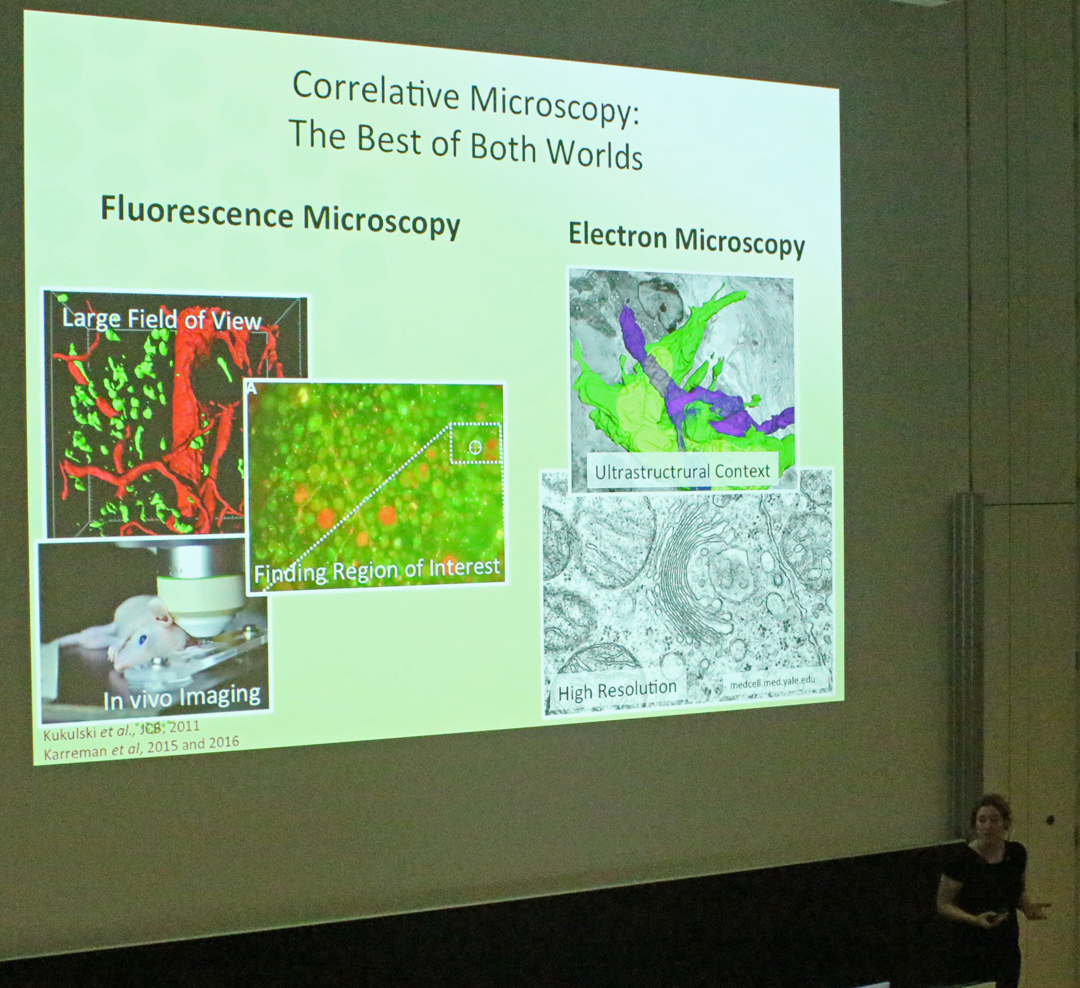
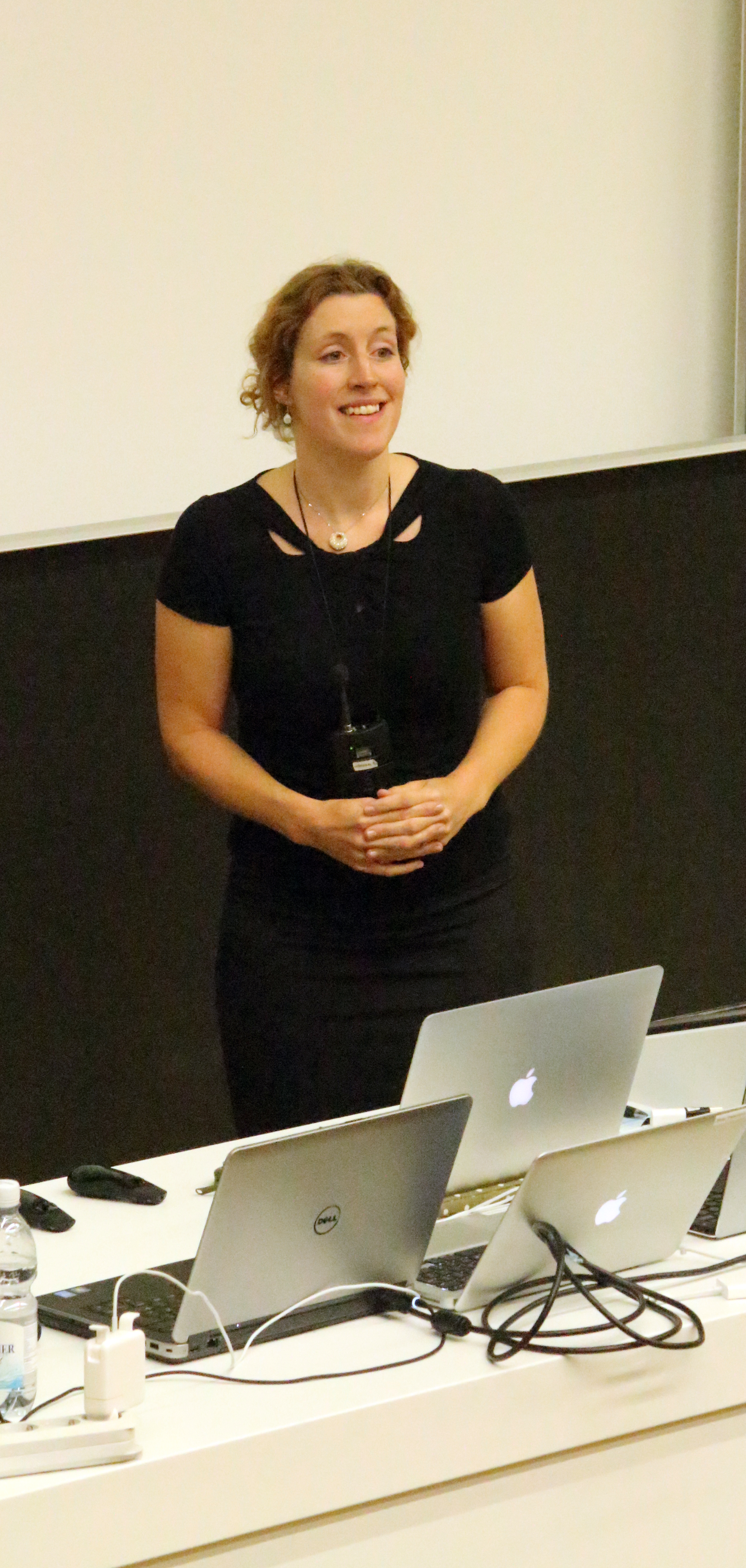
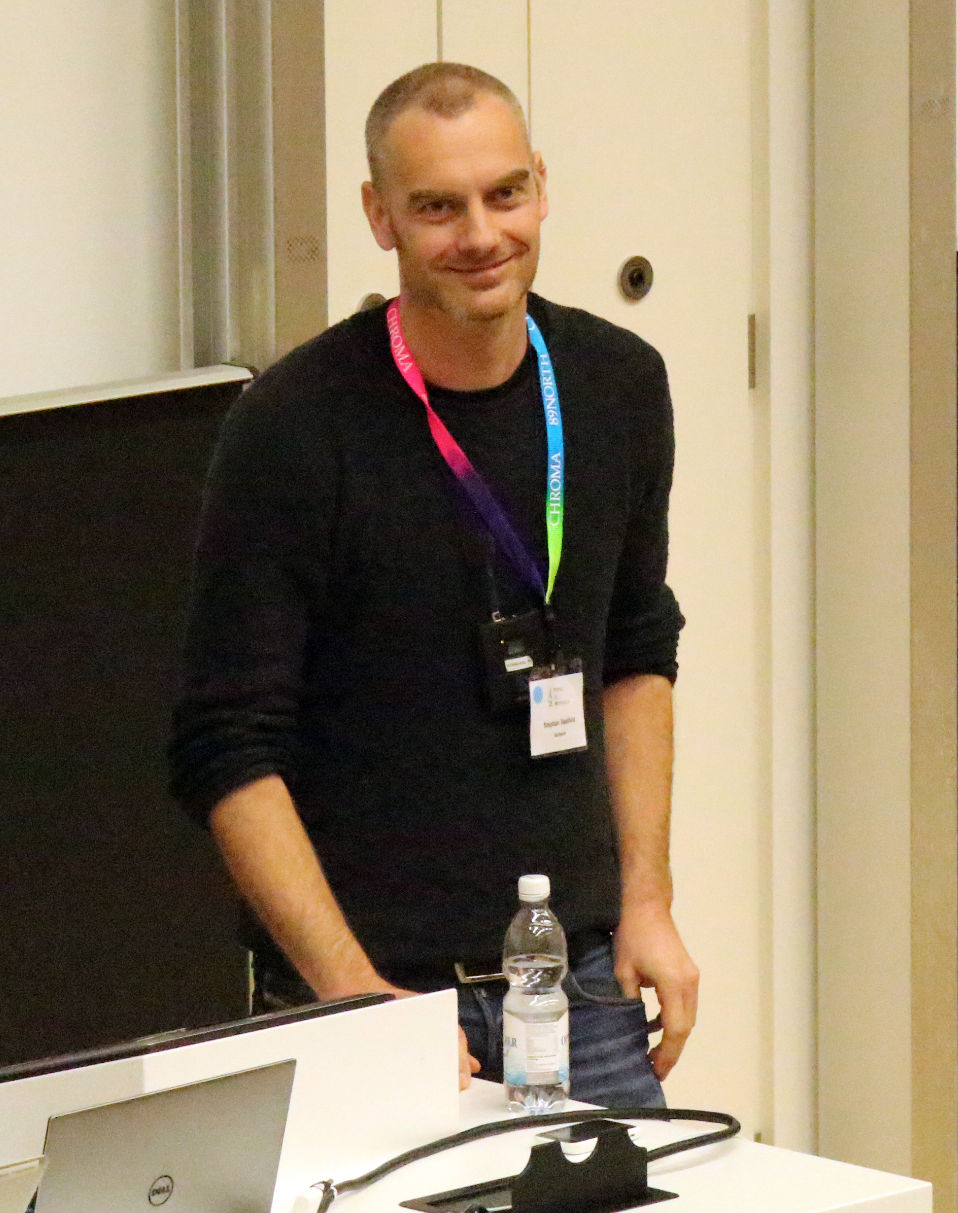
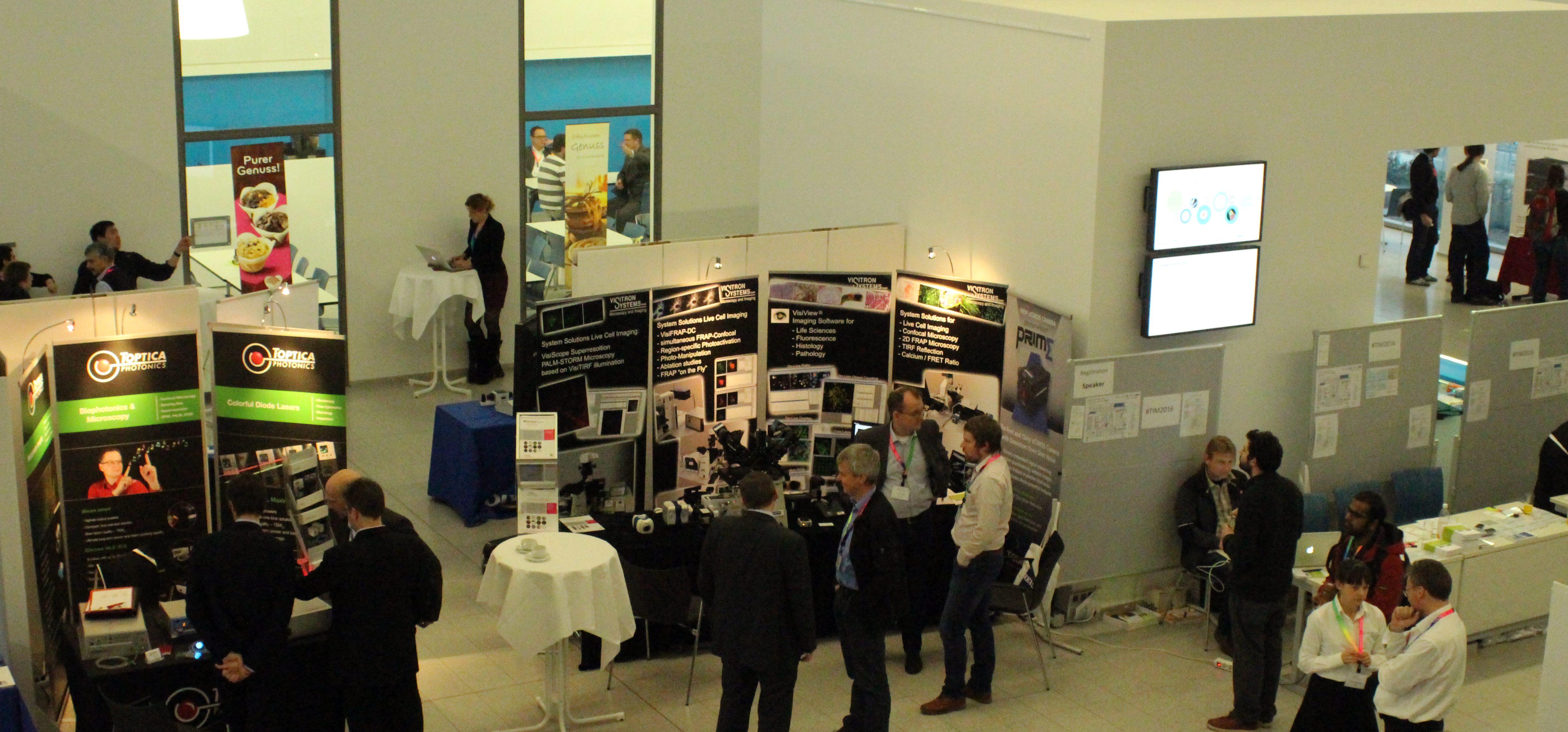
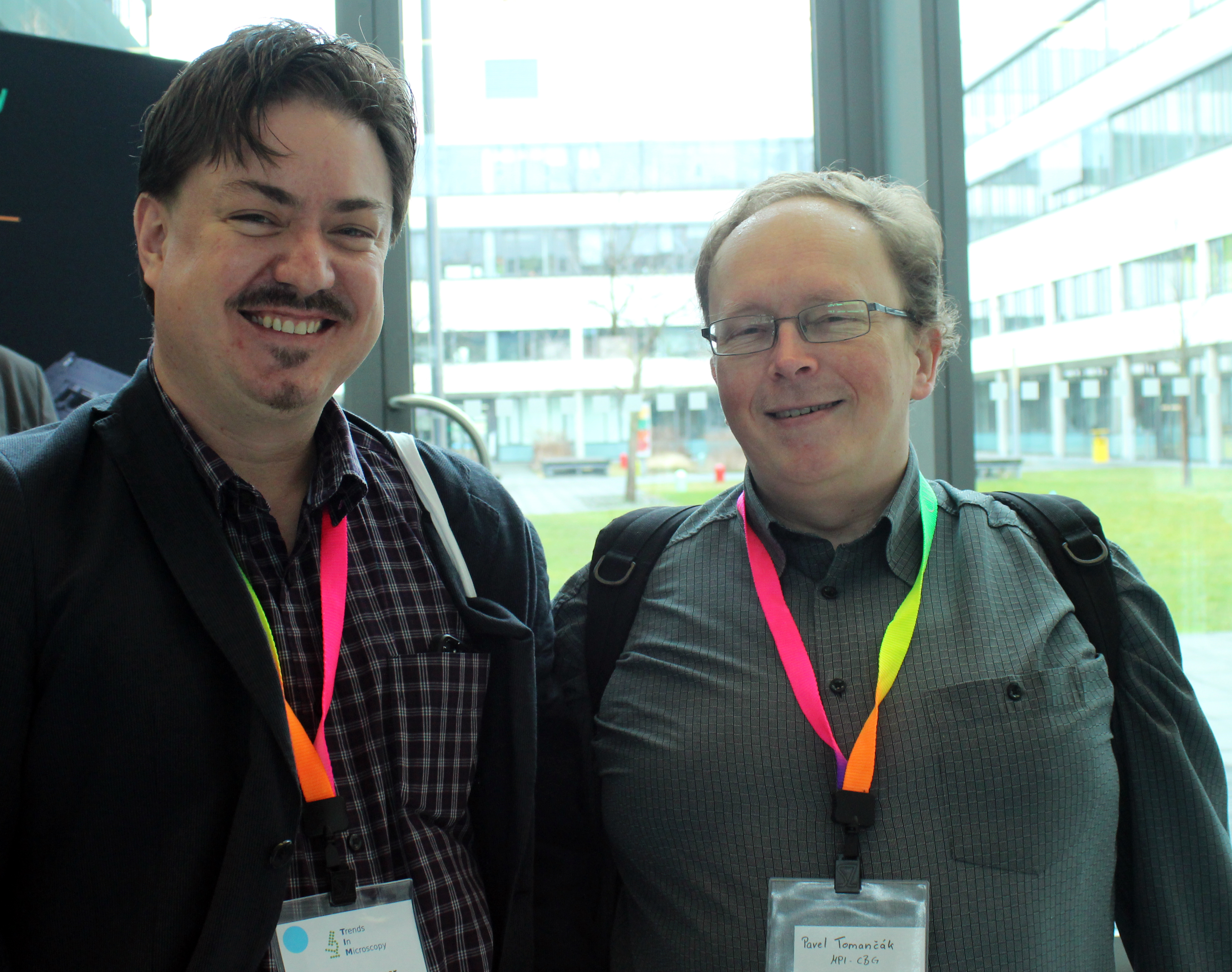
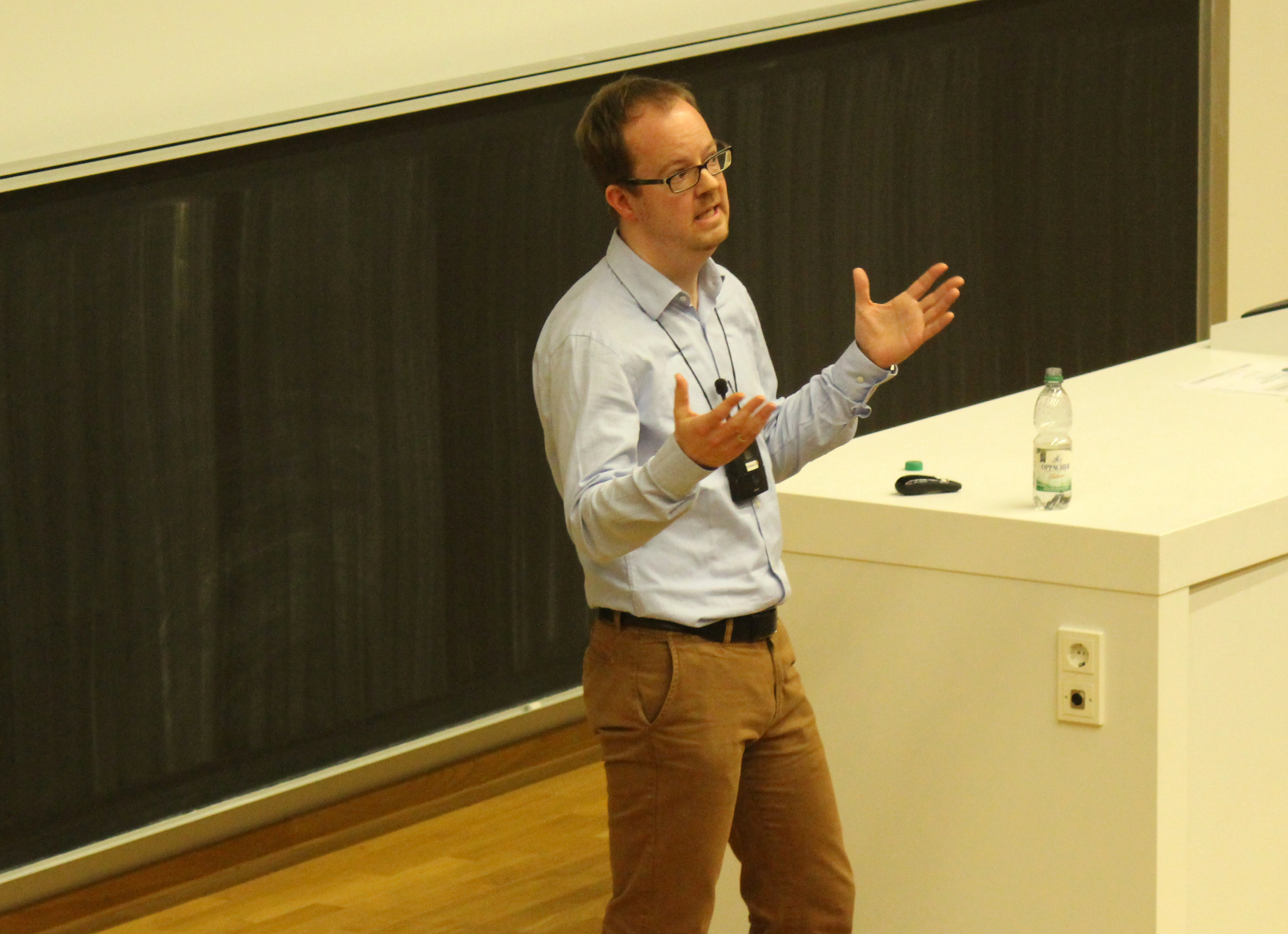
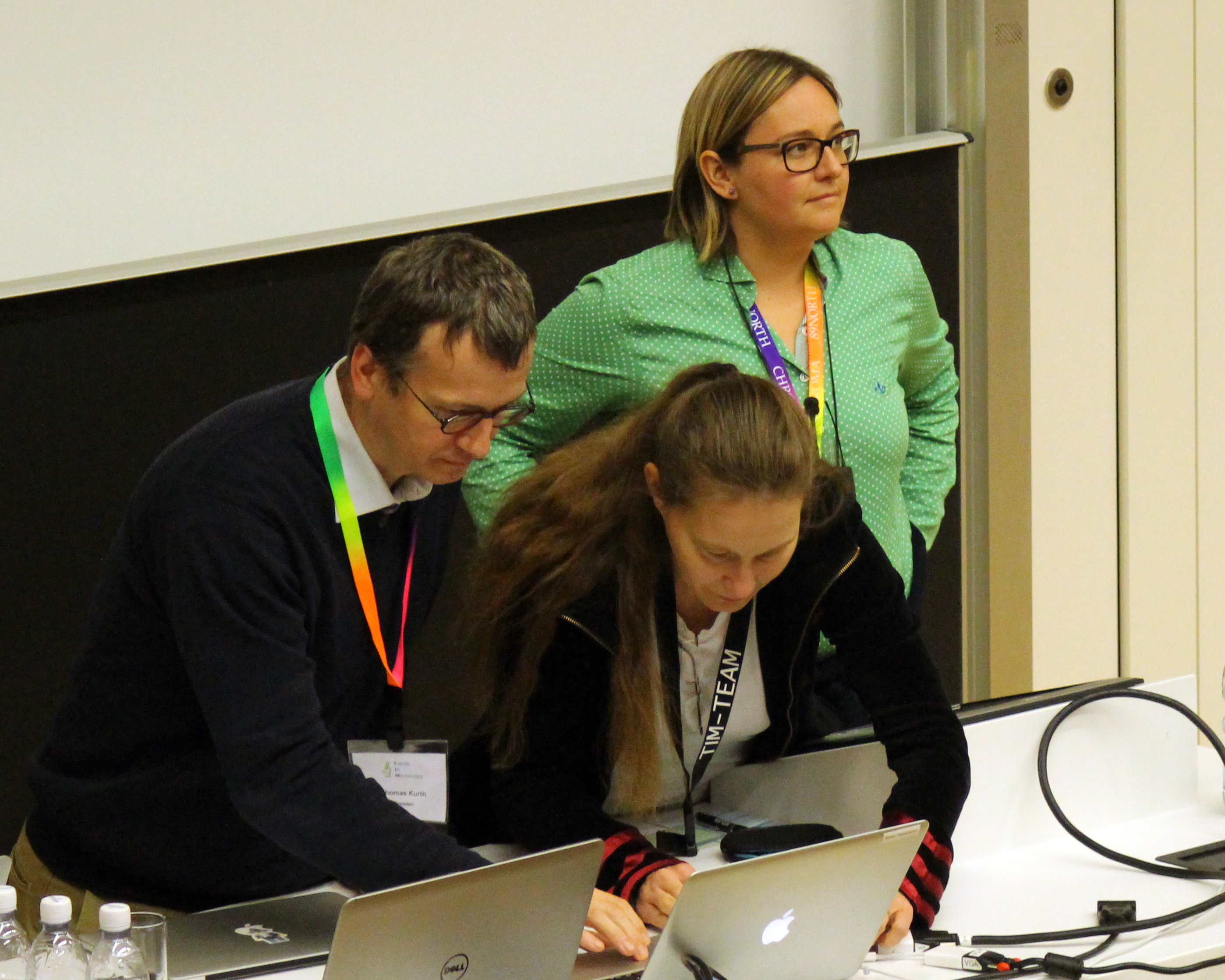
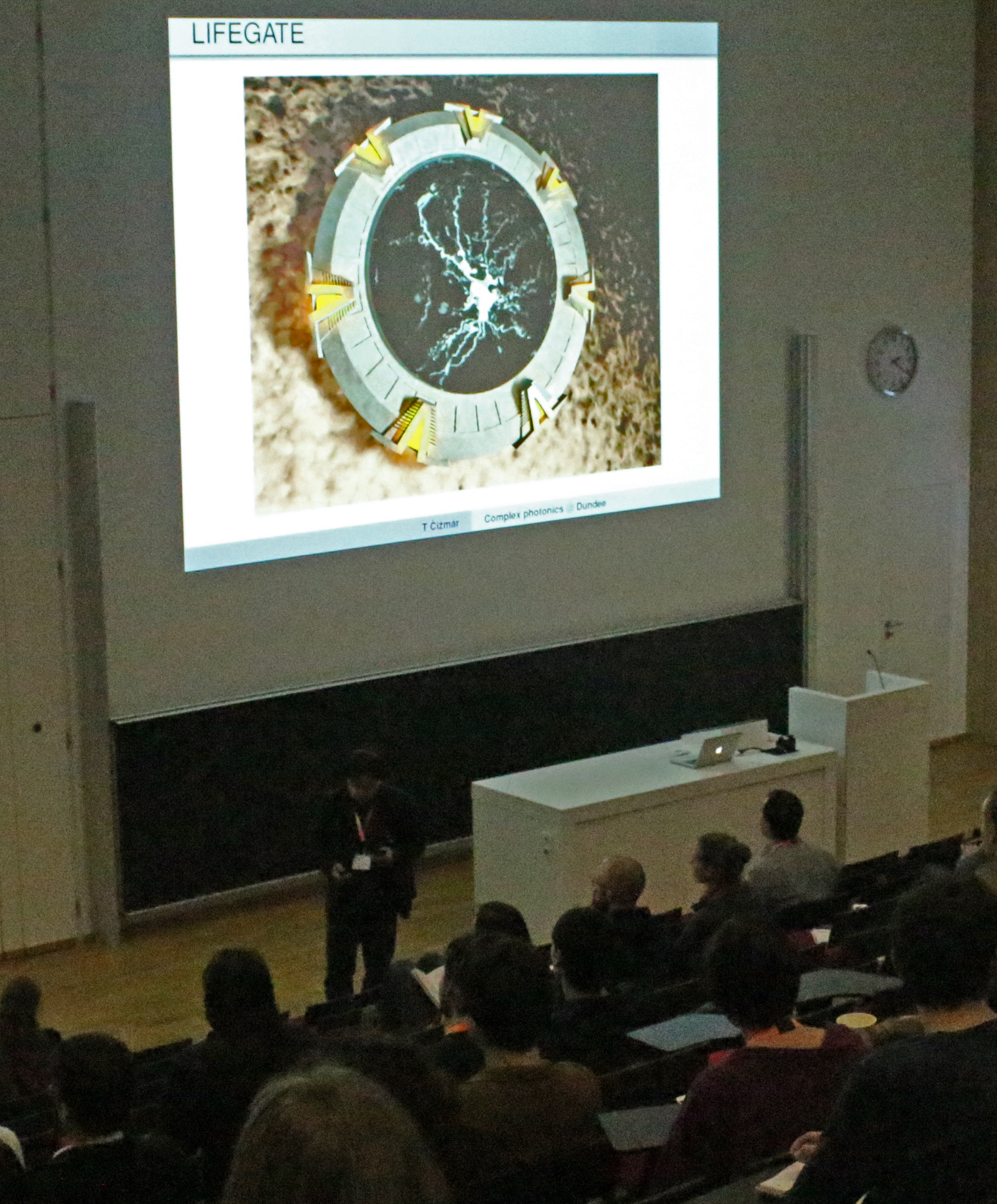
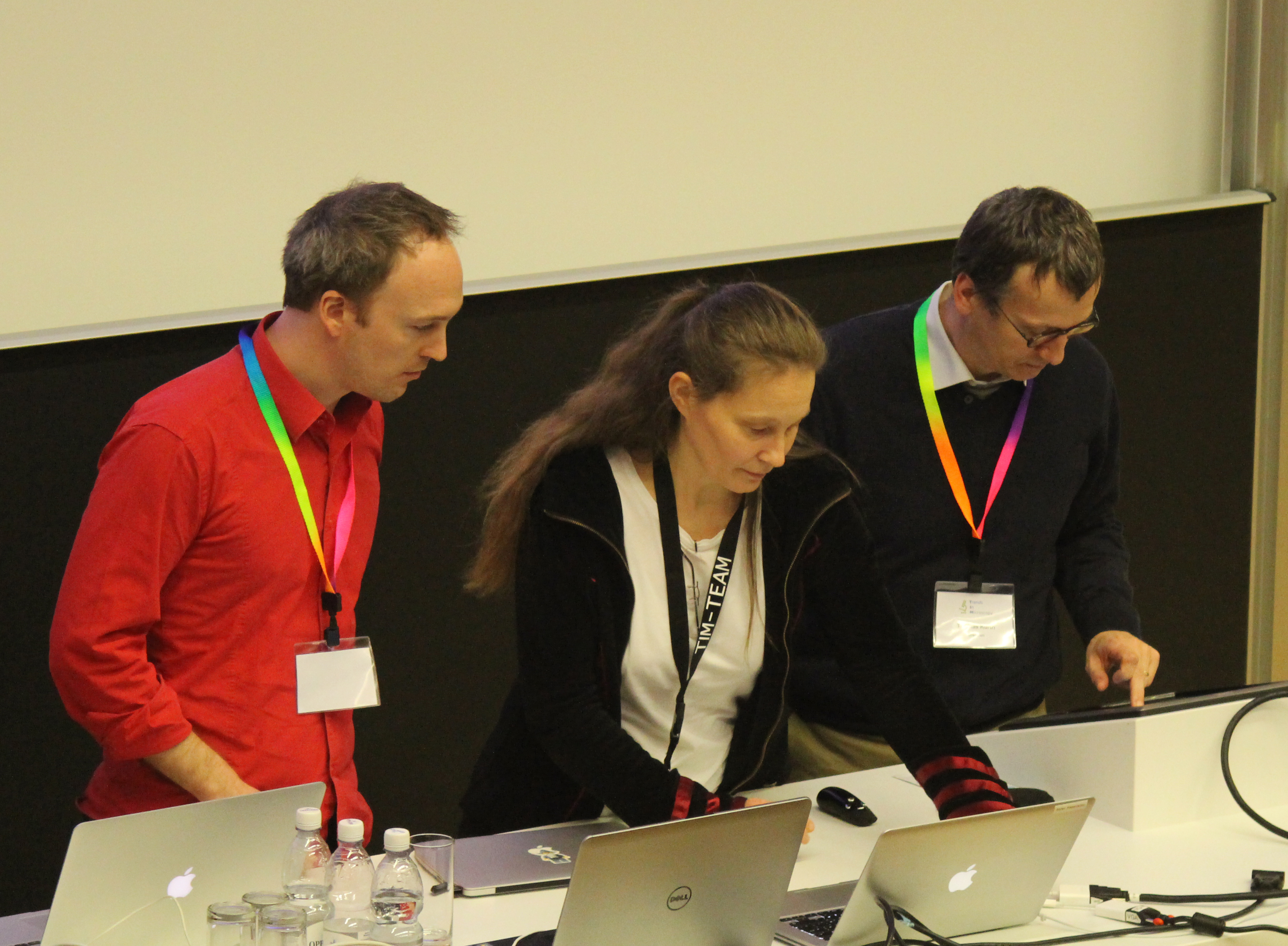
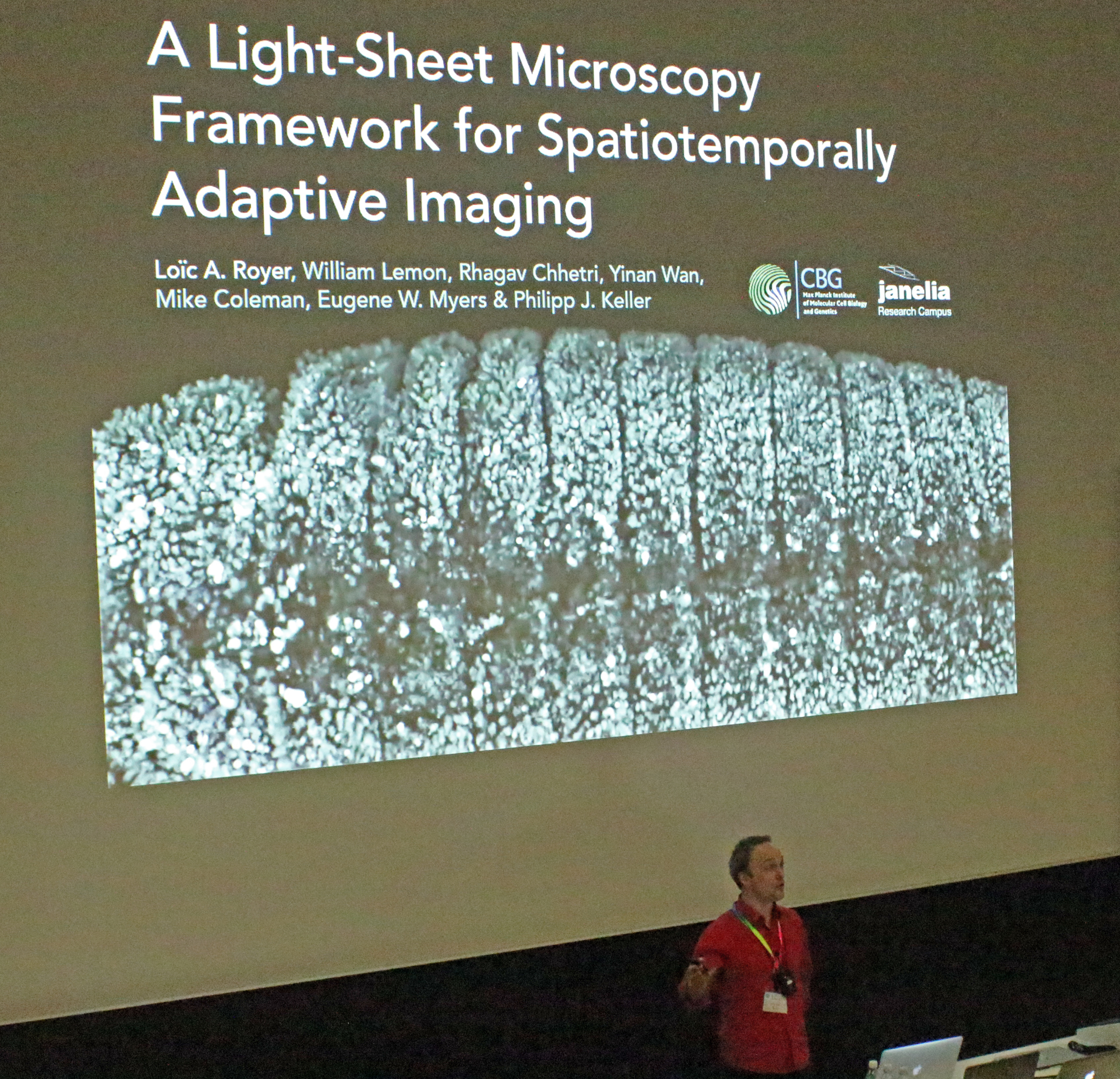
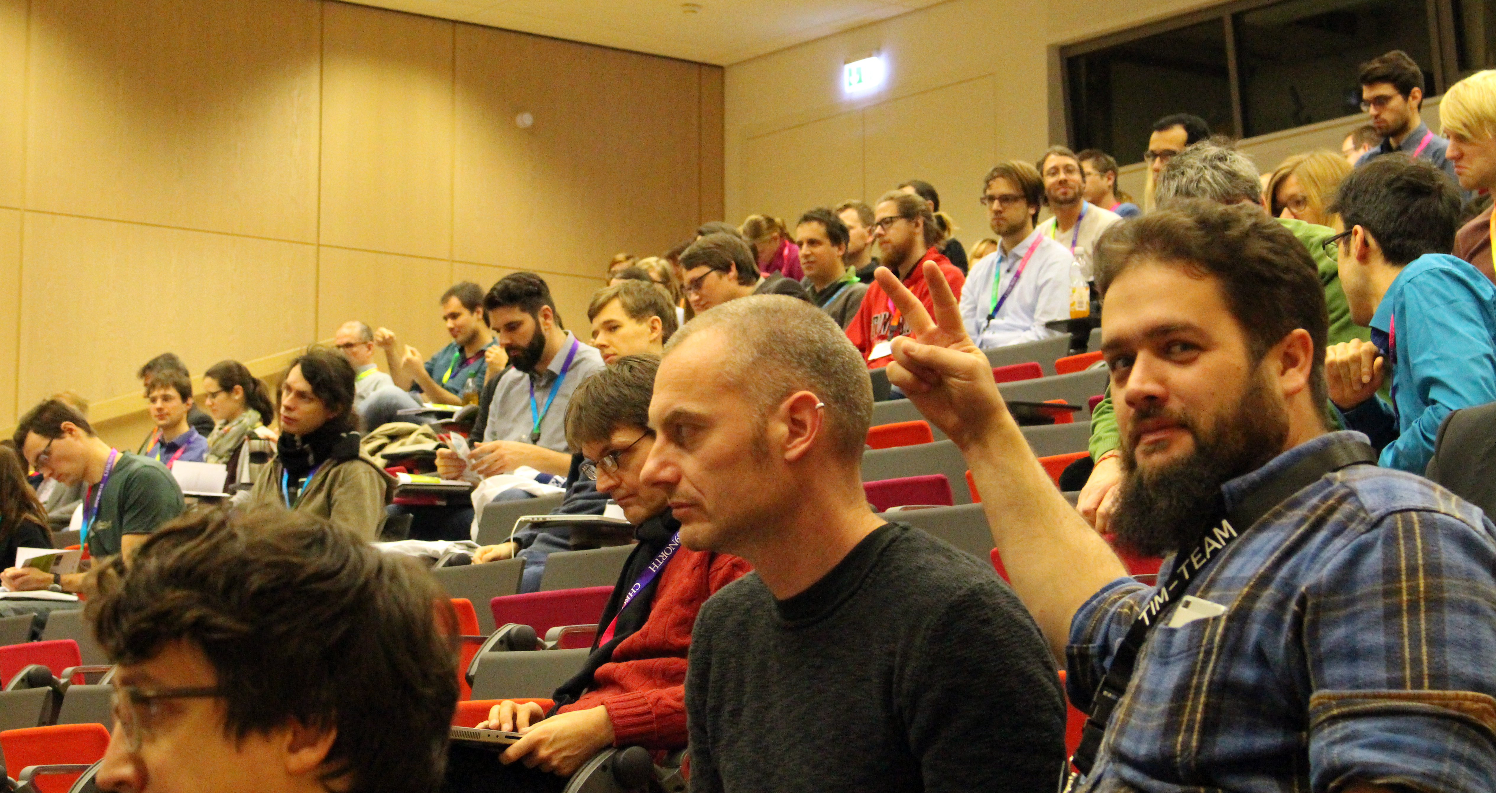
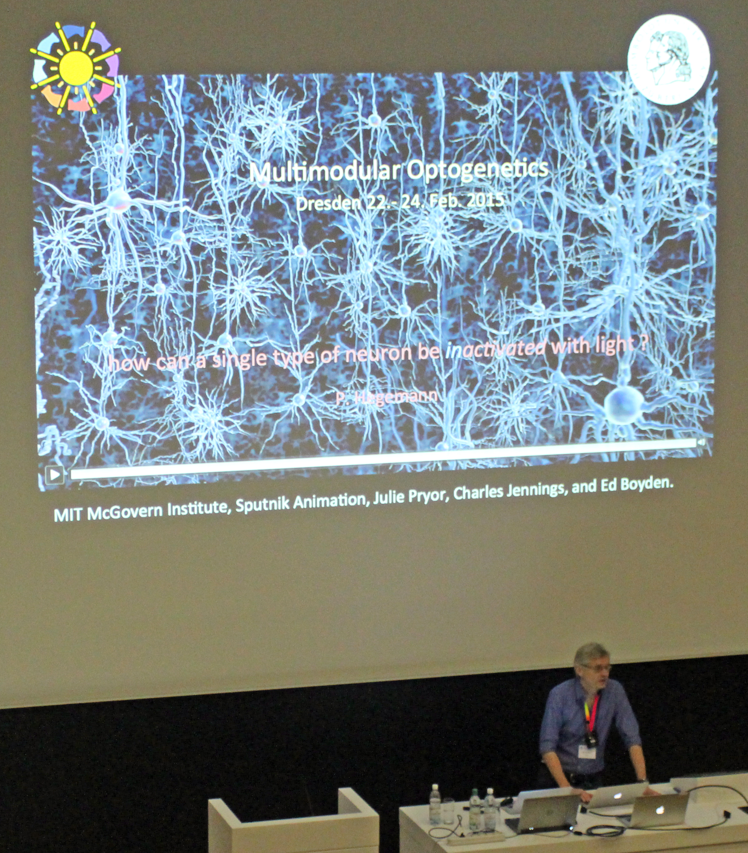
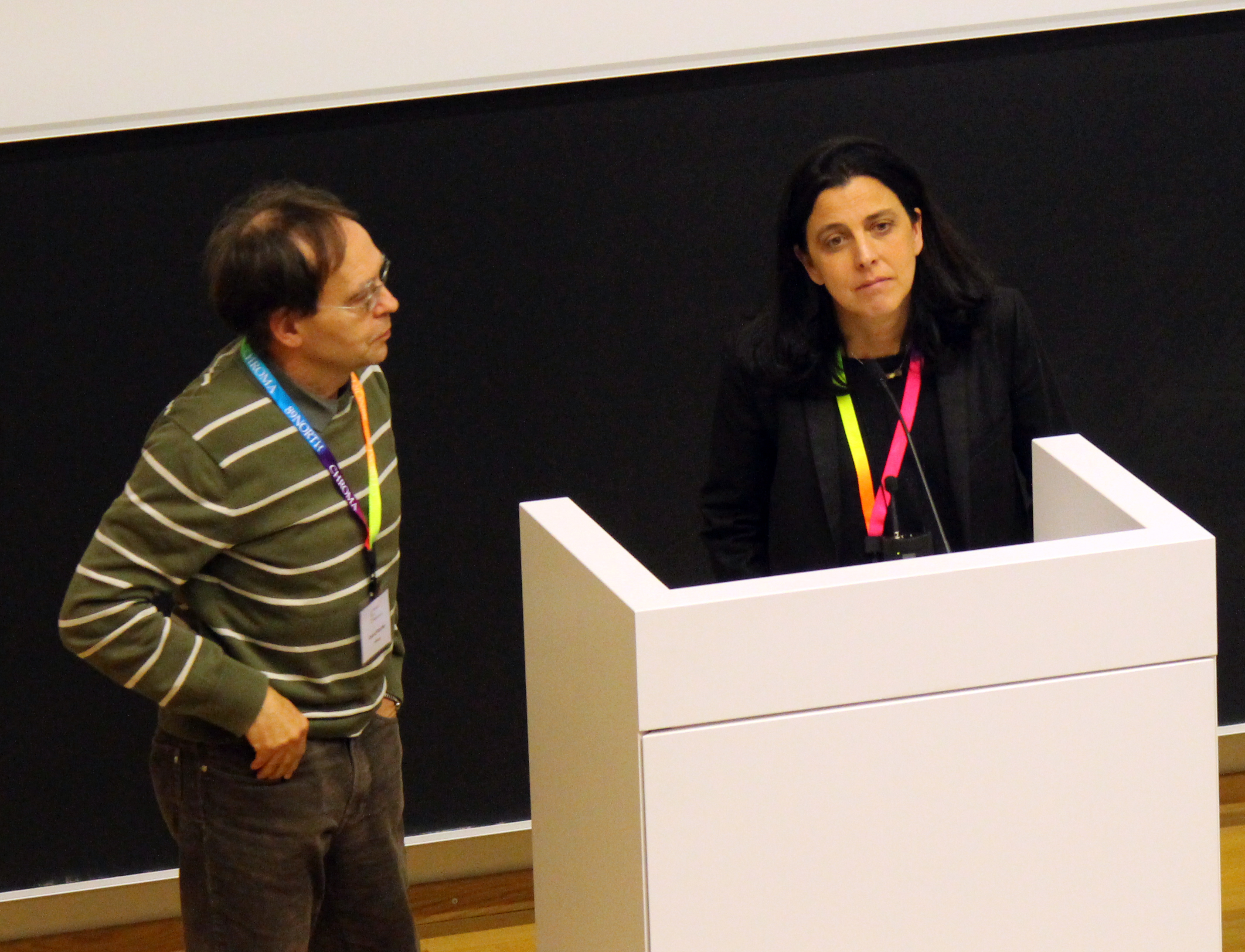
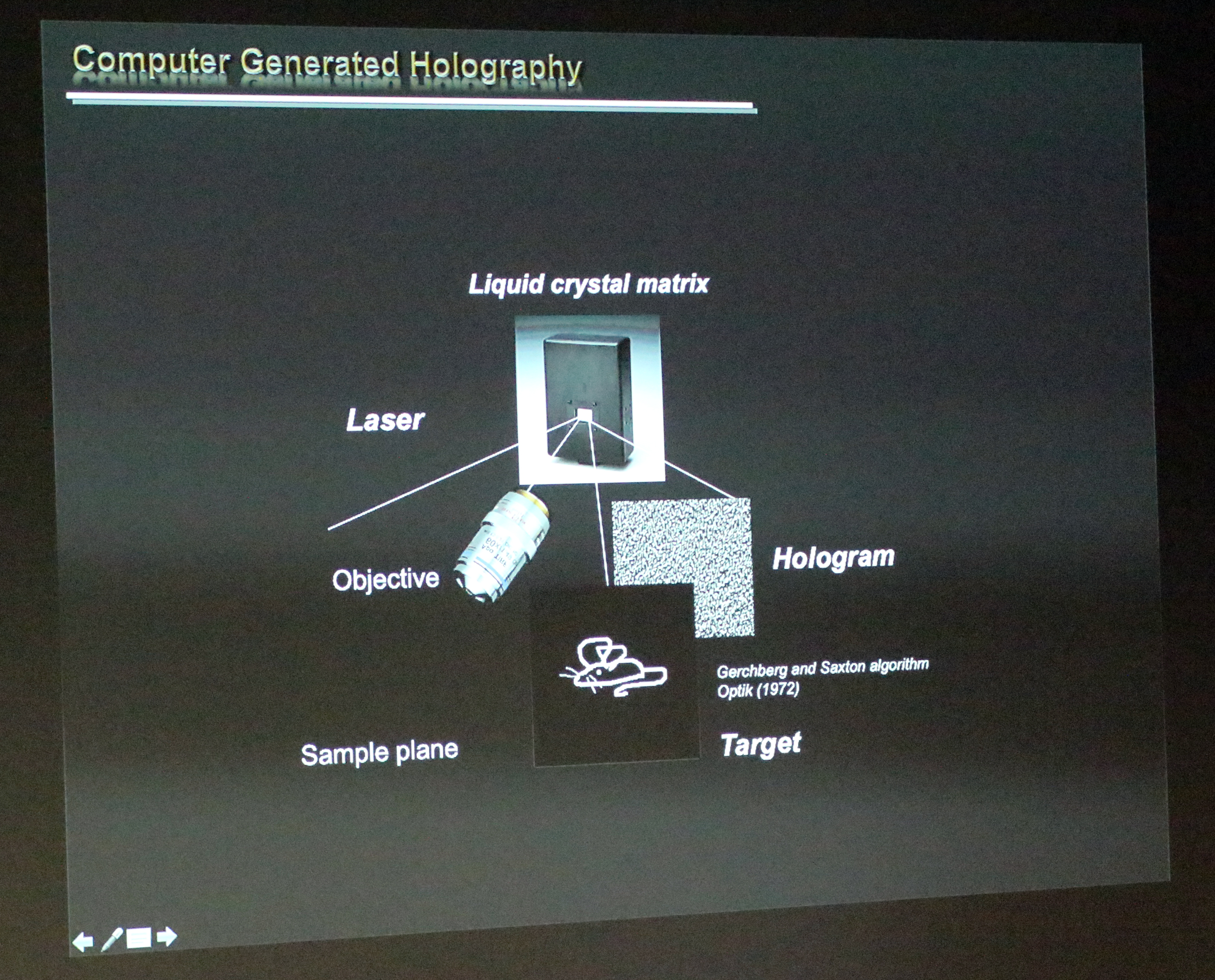
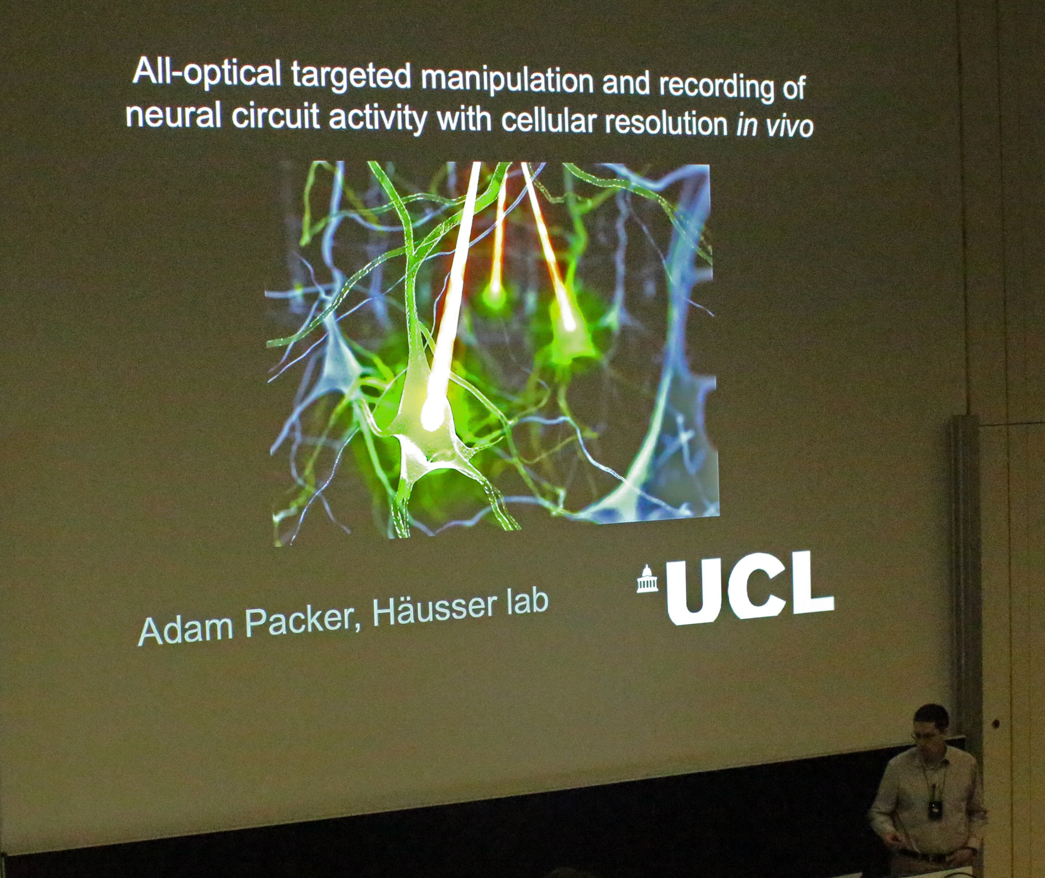
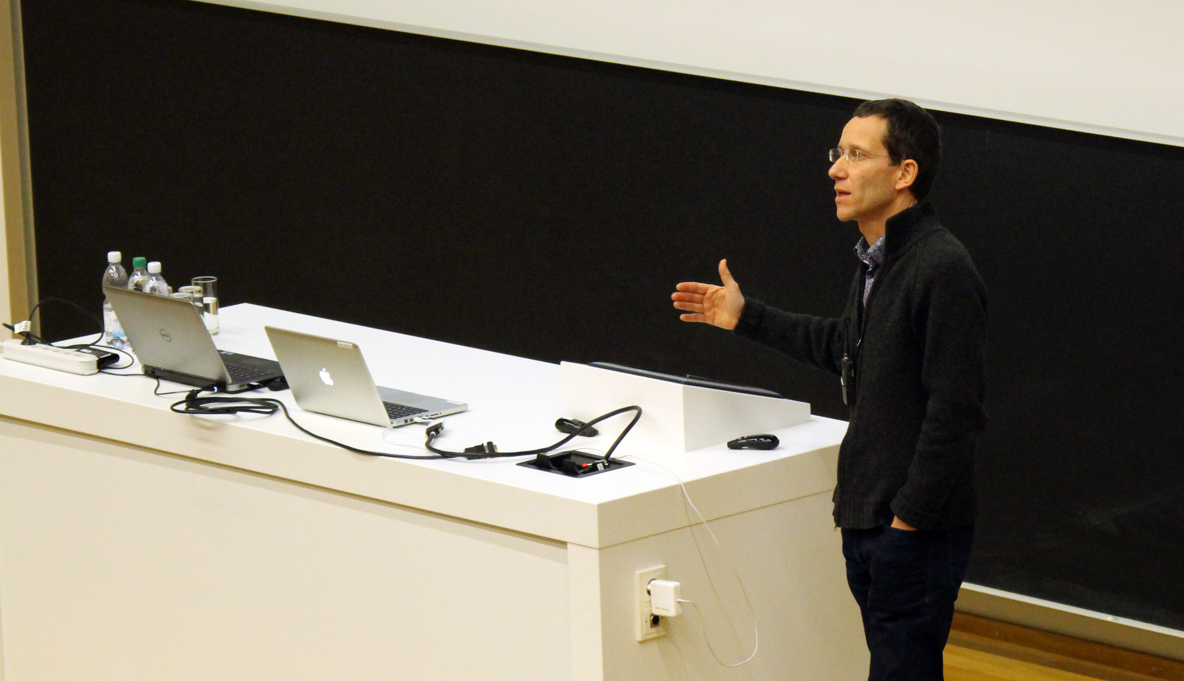
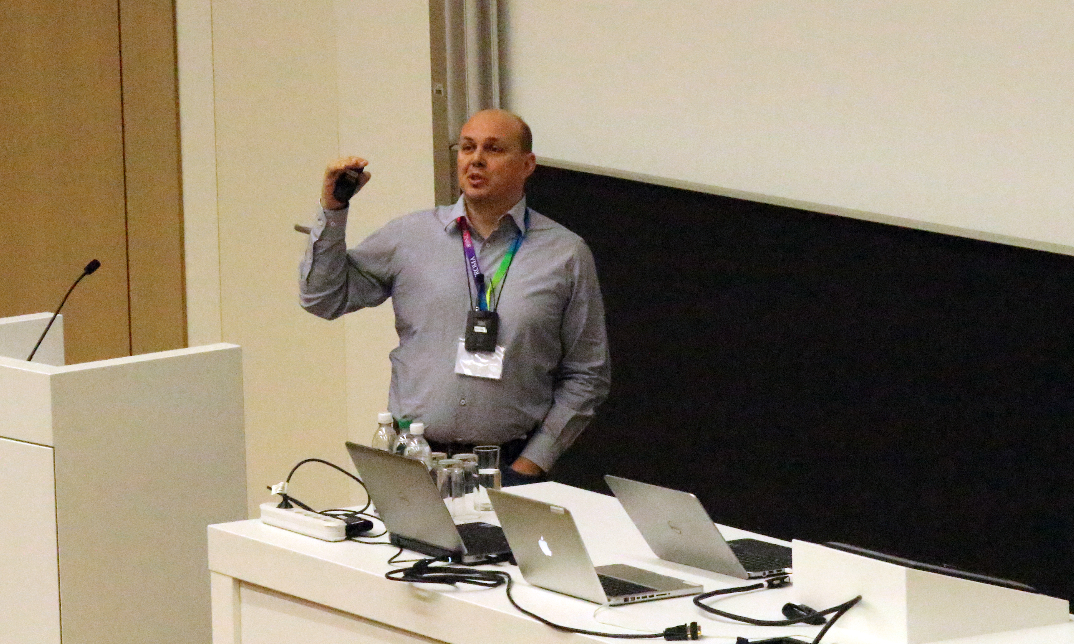
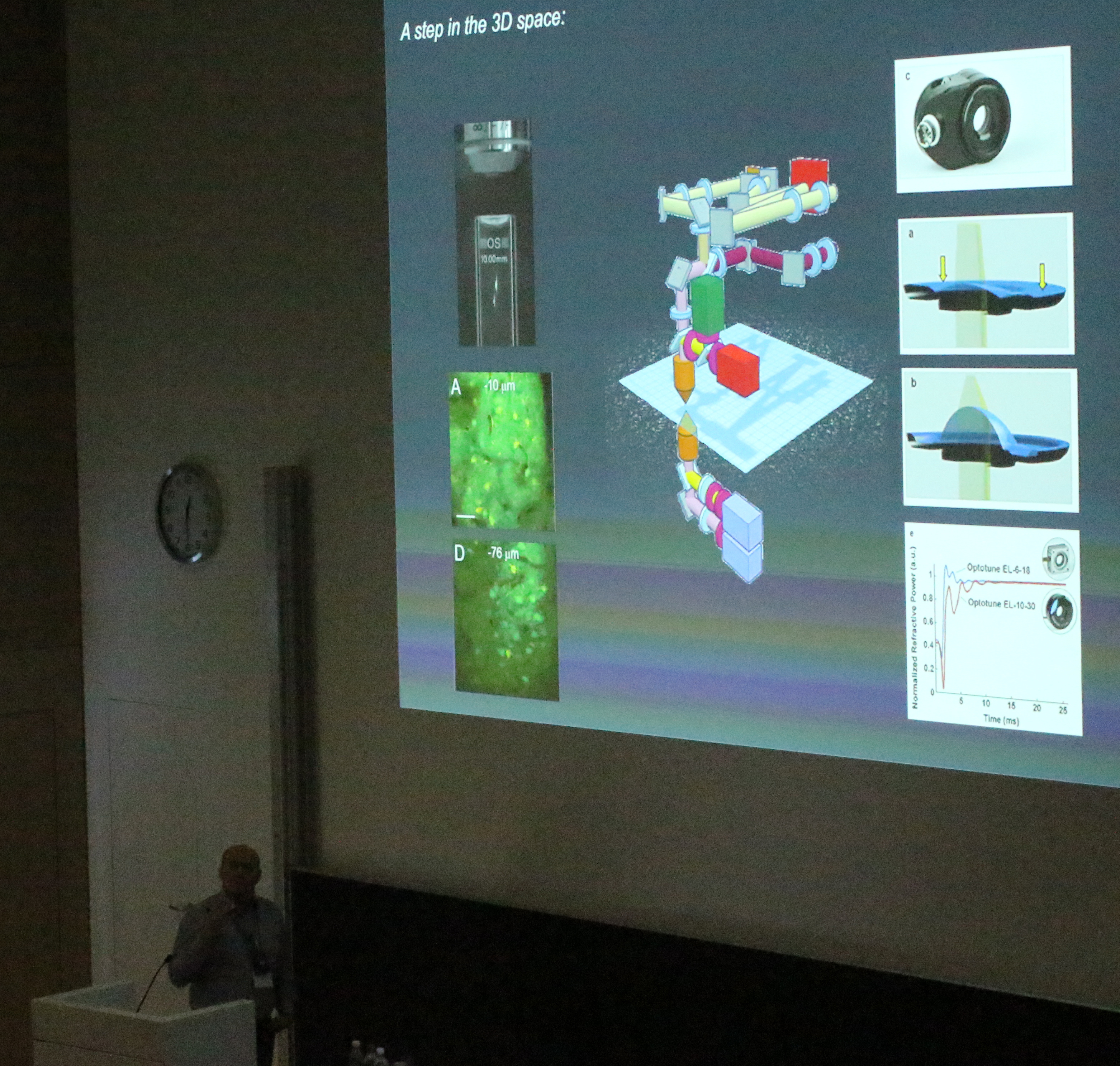
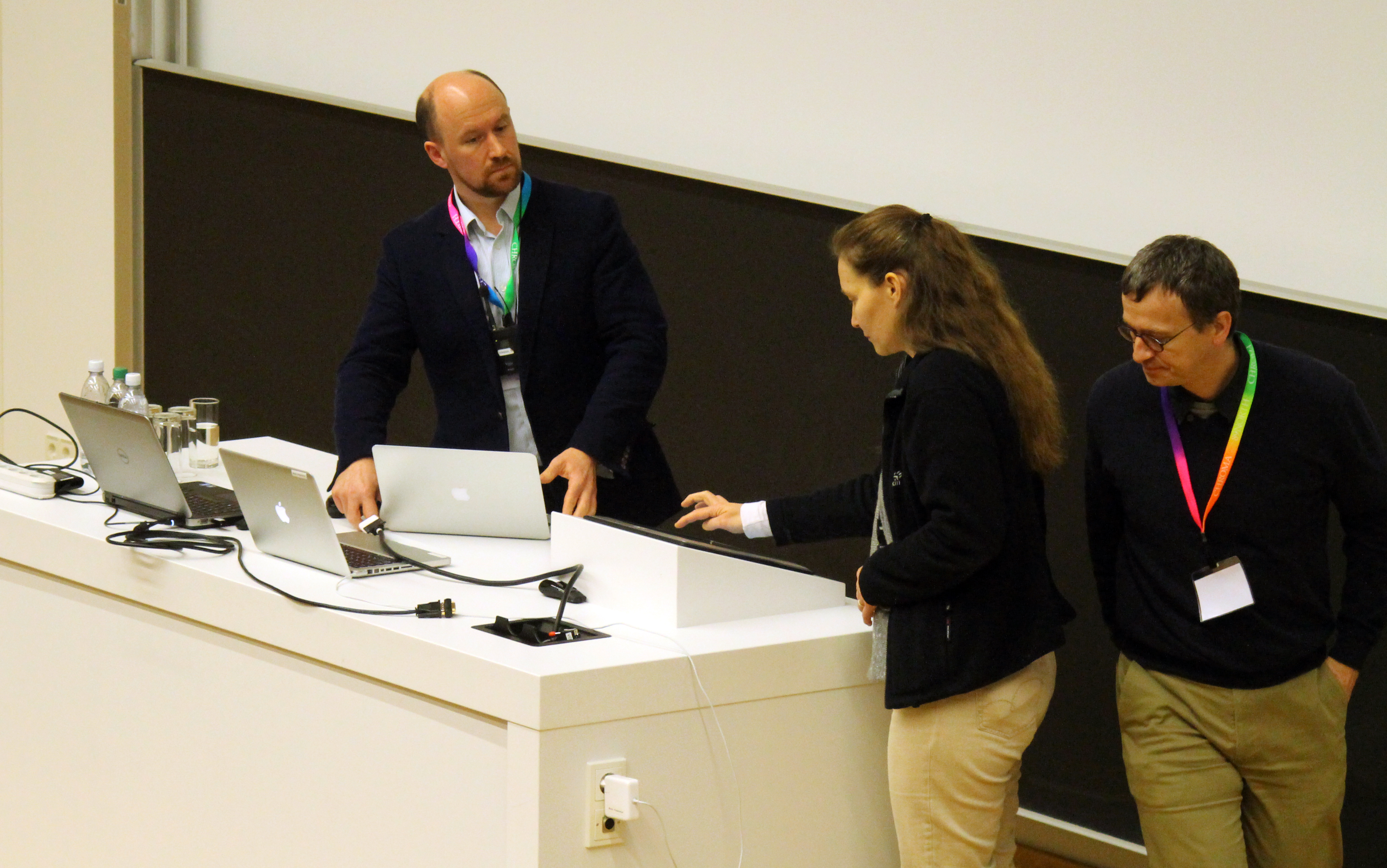
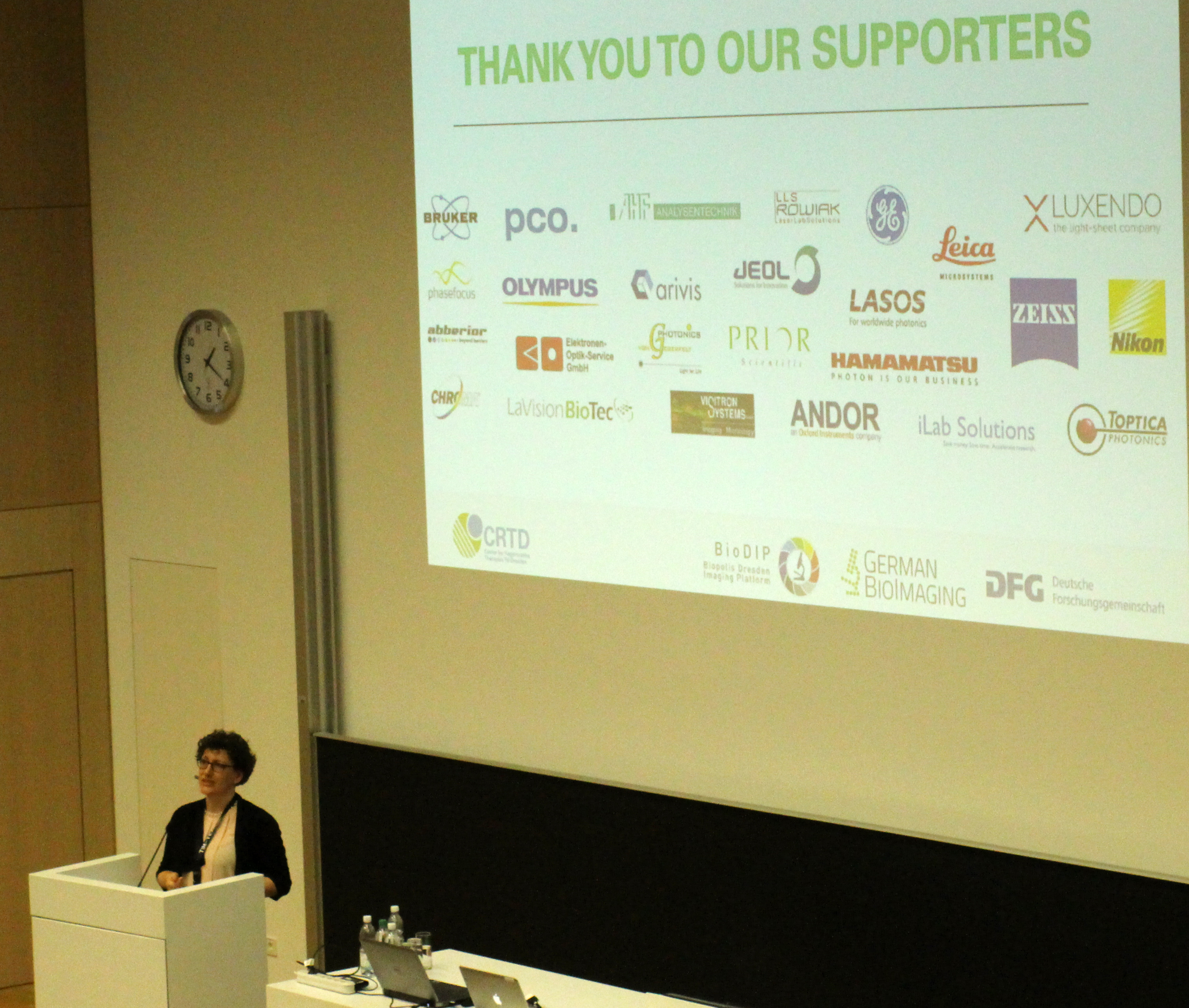
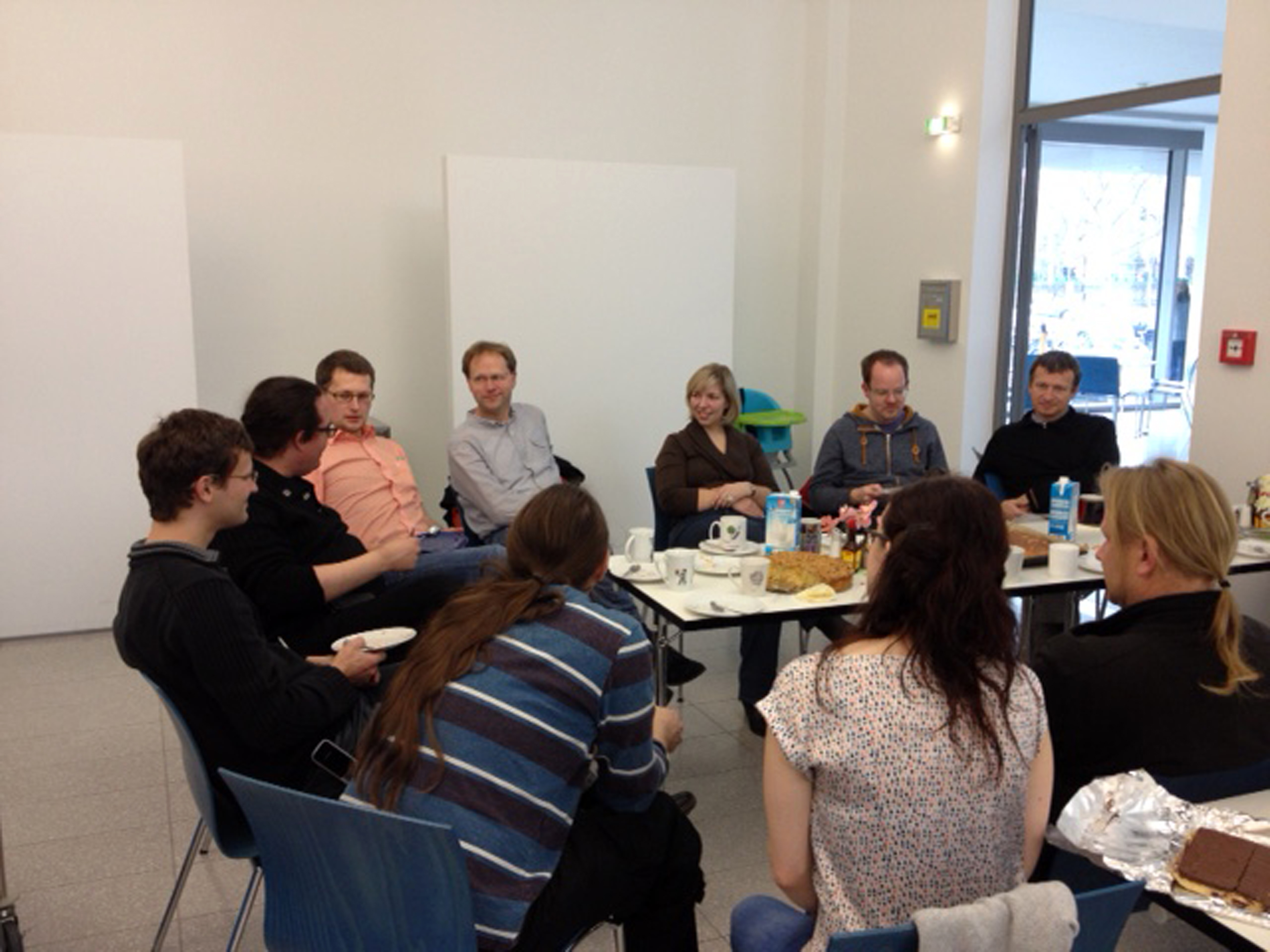
Conference Impressions of "Trends in Microscopy" 2016

TIM 2016 - Gerhard Rödel

Kick off - Gerhard Rödel, vice-rector for research at the TU Dresden opens "Trends in Microscopy" in 2016.

TIM 2016 - Michael Brand

Michael Brand, head of BioDIP welcomes all participants.

TIM 2016 - Elisa May

Elisa May, coordinator of GerBI in Konstanz welcomes all participants and starts the first session on Adaptive Optics.

TIM 2016 - Wolfgang Gässler

Wolfgang Gässler, an astronomer from Heidelberg, introduces the use of adaptive optics.

TIM 2016 - Audience

Attentive listeners

TIM 2016 - Na Ji

Na Ji, a group leader at Janelia Research Campus, develops optical methods for in vivo brain imaging.

TIM 2016 - Benjamin Judkewitz

Benjamin Judkewitz, professor of Bioimaging & Neurophotonics at the Charite/ Humboldt University in Berlin.

TIM 2016 - Peter Friedl

Peter Friedl employs various microscopic technolgies to reveal the mechanism of cancer invasion.

TIM 2016 - Allard Mosk

Allard P. Mosk, professor at the University of Twente, The Netherlands.

TIM 2016 - Helpers

Members of the TIM TEAM at the registration desks.

TIM 2016 - Moritz Kreysing

Moritz Kreysing, group leader at the MPI-CBG starts Session 2 on Phase & Holographic Imaging.

TIM 2016 - Achim Tieftrunk

Achim Tieftrunk welcomes all participants on behalf of the DFG which strongly supports this conference series.

TIM 2016 - Adam Wax

Adam Wax, Professor of Biomedical Engineering at Duke University, USA.

TIM 2016 - Gabriel Popescu

Gabriel Popescu, professor at the University of Illinois, USA works on quantitative phase imaging of cells and tissues.

TIM 2016 - Helpers

Britta and Thomas (front row) did a great job by assisting all speakers in technical matters. Robert was also memeber of the great TIM TEAM.

TIM 2016 - Mirjam Schürmann

Mirjam Schürmann, from the Guck lab in Dresden, after her talk.

TIM 2016 - Björn Kemper

Björn Kemper from the WWU in Münster talks about label-free digital holographic microscopy to image cells and tissue.

TIM 2016 - Alexander Jesacher

Alexander Jesacher, scientist at the Medical University in Innsbruck, presents a tunable diffractive/refractive hybrid lens.

TIM 2016 - Gaia Pigino

Gaia Pigino, group leader at the MPI-CBG, presents a nocel CLEM method.

TIM 2016 - Lucy Collison

Lucy Collison, researcher at the Francis Crick Institute, develops and applies correlative light and volume electron microscopy.

TIM 2016 - Julia Mahamid

Julia Mahamid,researcher at the MPI for Biochemistry after her talk.

TIM 2016 - Audience

Attentive listeners

TIM 2016 - Wanda Kukulski

Wanda Kukulski, group leader at the MRC Laboratory of Molecular Biology presented a hybrid imaging approach to dissect the mechanism of membrane remodelling.

TIM 2016 - Matthia Karreman

Matthia Karreman, researcher at the EMBL in Heidelberg uses multimodal correlative microscopy to image single tumor cells in vivo.

TIM 2016 - Matthia Karreman

Matthia Karreman answering questions after her talk.

TIM 2016 - Stephan Saalfeld

Stephan Saalfeld, group leader at Janelia Research Campus, talked about image registration and interpretation of compromised data from complementary imaging modalities.

TIM 2016 - Exhibitors

More than 20 exhibitors were present during the meeting.

TIM 2016 - Tomas Cizmar

Tomas Cizmar and Pavel Tomancak enjoy the coffee break.

TIM 2016 - Jan Huisken

Jan Huisken, group leader at the MPI-CBG presents his idea of a smart microscope.

TIM 2016 - Vera Hirschfeld-Warneken

Vera Hirschfeld-Warneken from Stuttgart is set up by Britta & Thomas for her presentation on the application of reflective interference contrast microscopy.

TIM 2016 - Tomas Cizmar

Tomas Cizmar,researcher at the University of Dundee presented the use of multimode waveguides as fibre-based endoscopes for imaging deep inside living

TIM 2016 - Loic Royer

Loic Royer, researcher at the MPI-CBG is set up for his presentation by Britta and Thomas

TIM 2016 - Loic Royer

Loic Royer begins his talk on multimodal light shet microscopy.

TIM 2016 - Audience

Naughty Pete …

TIM 2016 - Peter Hegemann

Peter Hegemann, professor at the Humboldt University in Berlin, gives an introduction to optogenetics.

TIM 2016 - Valentina Emiliani

Valentina Emiliani, director of the Neurophotonics Laboratory in Paris, employs multi-plane illumination technology to stimulate several single neurons simultaneously in 3D.

TIM 2016 - Valentina Emiliani

Computer generated holography.

TIM 2016 - Adam Packer

Adam Packer, researcher at UCL in London, talks about imaging and manipulation of neural circuit activity.

TIM 2016 - Thomas Oertner

Thomas Oertner, Professor at the University of Hamburg, after his presentation.

TIM 2016 - Marco dal Maschio

Marco dal Maschio, researcher at the MPI for Neurobiology, presents approaches to combine imaging and photo-stimulation of brain circuits.

TIM 2016 - Marco dal Maschio

Marco dal Maschio during his presentation.

TIM 2016 - Martin Booth

Martin Booth, professor of engineering at the University of Oxford, is set up by Britta and Thomas for his presentation on adaptive optics for fluorescent nanoscopy.

TIM 2016 - Roundup

Roundup: Hella says thank you to all participants, speakers, supporters & helpers and closes the meeting.

TIM 2016 -Thank You!

We thank our local helpers and supporters with home-made cake.

TIM 2016 - Thank You!

We thank our local helpers and supporters with home-made cake.

TIM 2016 - Thank You!

We thank our local helpers and supporters with home-made cake.

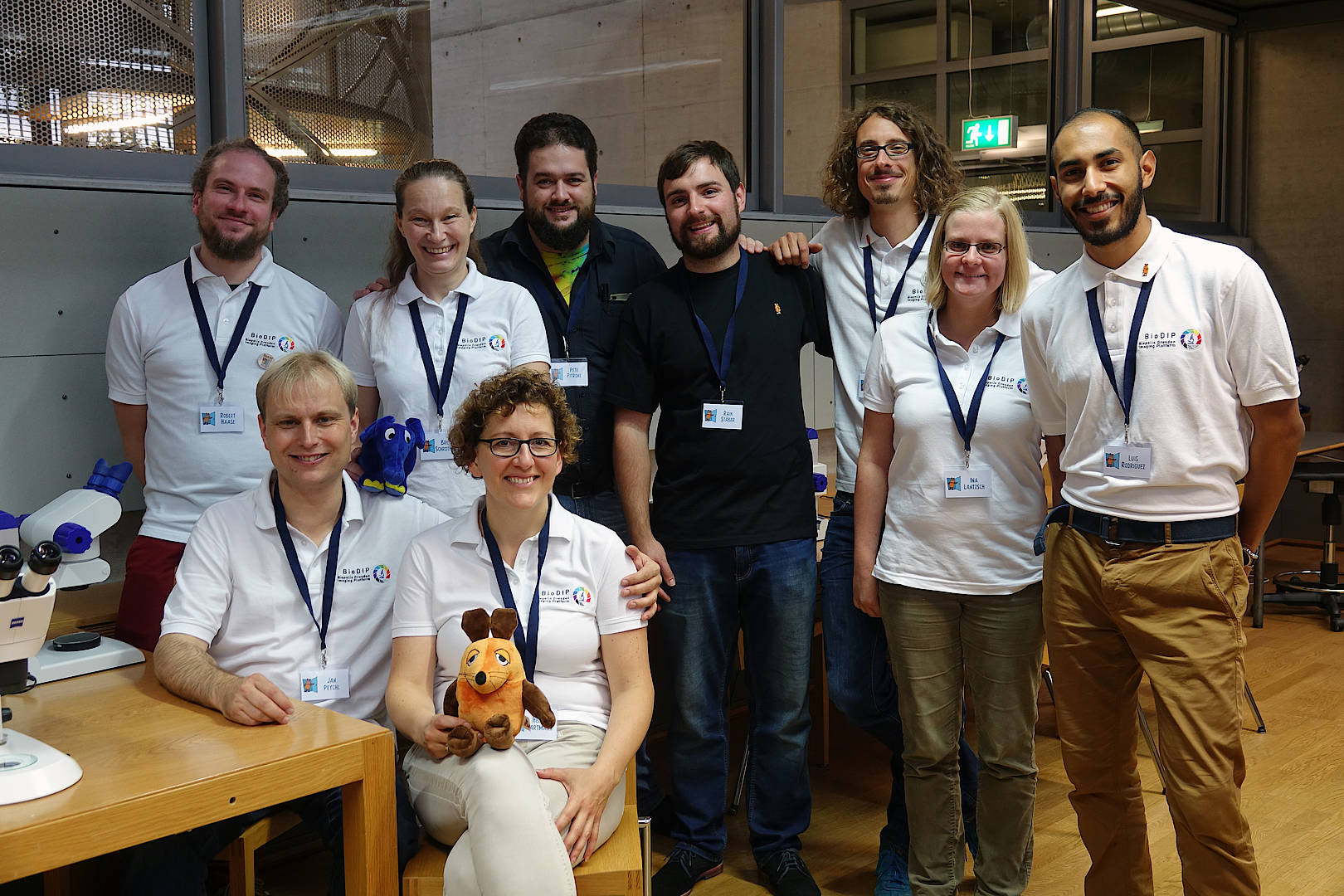
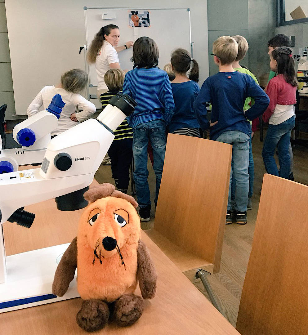
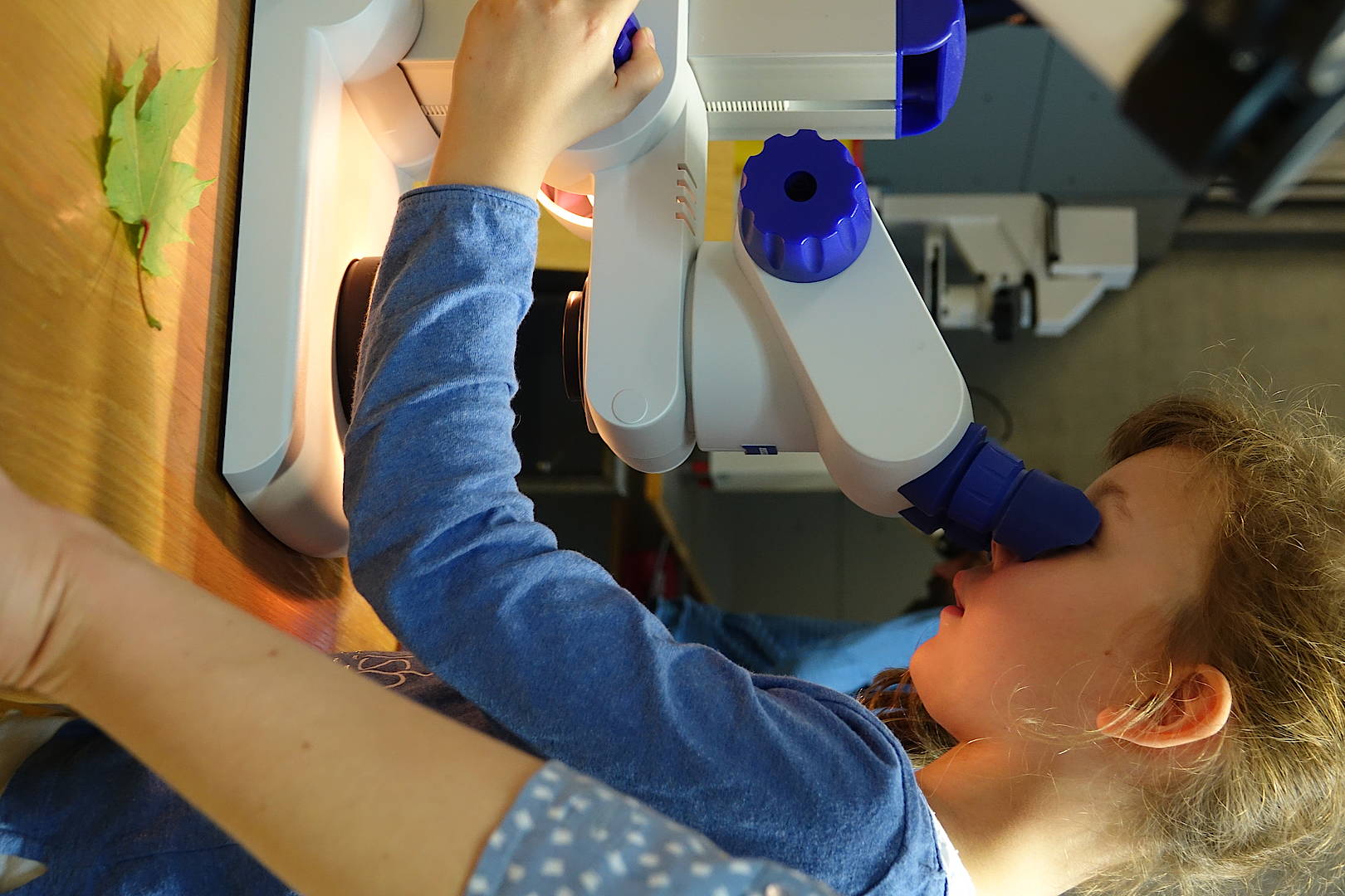
Maustüröffner Tag 2017

BioDIP team and friends in the end of the day.