Electron microscopy of the amphibian model systems Xenopus laevis and Ambystoma mexicanum.
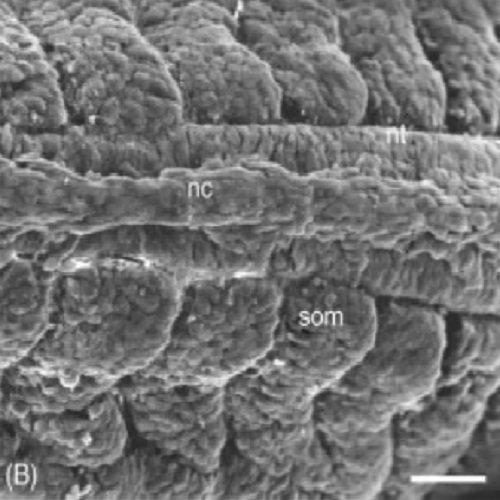
In this chapter we provide a set of different protocols for the ultrastructural analysis of amphibian (Xenopus, axolotl) tissues, mostly of embryonic origin. For Xenopus these methods include: (1) embedding gastrulae and tailbud embryos into Spurr's resin for TEM, (2) post-embedding labeling of methacrylate (K4M) and cryosections through adult and embryonic epithelia for correlative LM and TEM, and (3) pre-embedding labeling of embryonic tissues with silver-enhanced nanogold. For the axolotl (Ambystoma mexicanum) we present the following methods: (1) SEM of migrating neural crest (NC) cells; (2) SEM and TEM of extracellular matrix (ECM) material; (3) Cryo-SEM of extracellular matrix (ECM) material after cryoimmobilization; and (4) TEM analysis of hyaluronan using high-pressure freezing and HABP labeling. These methods provide exemplary approaches for a variety of questions in the field of amphibian development and regeneration, and focus on cell biological issues that can only be answered with fine structural imaging methods, such as electron microscopy.

- Methods Cell Biol. 2010;96:395-423
- 2010
- Imaging Technologies Development
- 20869532
- PubMed
Enabled by:
