Automated tracing of microtubules in electron tomograms of plastic embedded samples of Caenorhabditis elegans embryos.
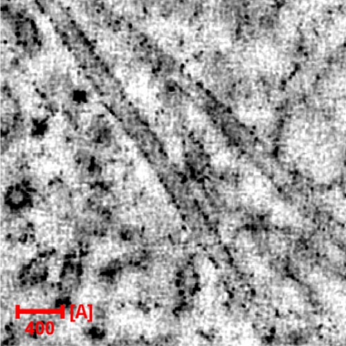
The ability to rapidly assess microtubule number in 3D image stacks from electron tomograms is essential for collecting statistically meaningful data sets. Here we implement microtubule tracing using 3D template matching. We evaluate our results by comparing the automatically traced centerlines to manual tracings in a large number of electron tomograms of the centrosome of the early Caenorhabditis elegans embryo. Furthermore, we give a qualitative description of the tracing results for three other types of samples. For dual-axis tomograms, the automatic tracing yields 4% false negatives and 8% false positives on average. For single-axis tomograms, the accuracy of tracing is lower (16% false negatives and 14% false positives) due to the missing wedge in electron tomography. We also implemented an editor specifically designed for correcting the automatic tracing. Besides, this editor can be used for annotating microtubules. The automatic tracing together with a manual correction significantly reduces the amount of manual labor for tracing microtubule centerlines so that large-scale analysis of microtubule network properties becomes feasible.

- J. Struct. Biol. 2012 May 13;178(2):129-38
- 2012
- Imaging Technologies Development
- 22182731
- PubMed
Enabled by:
