Links
From BioDIP
(Difference between revisions)
(→Objective Data) |
|||
| (19 intermediate revisions by one user not shown) | |||
| Line 52: | Line 52: | ||
* [http://www.nikon.com/products/instruments/lineup/bioscience/biological-microscopes/accessory/objectives/index.htm Nikon objective list] | * [http://www.nikon.com/products/instruments/lineup/bioscience/biological-microscopes/accessory/objectives/index.htm Nikon objective list] | ||
| − | + | * [[media:Objective specifications.jpg|Objective specifications]] | |
| + | === Refractive Index=== | ||
| + | Measurements were performed at MPI-CBG LMF, with the J457 Automatic Refractometer (RUDOPLH) for each medium, in a temperature range between 10 and 60 Celsius degrees at 1 degree steps. | ||
* [[media:Immersion oil.pdf|Immersion Oil]] | * [[media:Immersion oil.pdf|Immersion Oil]] | ||
* [[media:Immersion water.pdf|Immersion Water]] | * [[media:Immersion water.pdf|Immersion Water]] | ||
| − | * [[media: | + | * [[media:Silicone.pdf|Silicone Immersion Oil]] |
| − | * [[media: | + | * [[media:Glycerol type G leica.pdf|Glycerol Type G]] |
* [[media:Glycerol 100%.pdf|Glycerol 100%]] | * [[media:Glycerol 100%.pdf|Glycerol 100%]] | ||
| + | |||
| + | * [[media:Glycerol 50%.pdf|Glycerol 50%]] | ||
| + | |||
| + | * [[media:Ultrasonic Gel.pdf|Ultrasonic Gel]] | ||
| + | |||
| + | * [[media:Vidisic.pdf|Vidisic]] | ||
== Free Software == | == Free Software == | ||
Latest revision as of 13:17, 13 October 2015
Contents |
[edit] Microscopy - Infos and Introductions

- Molecular Expressions Microscopy Primer
very comprehensive - provides information about various microscopy techniques, stainings and much more- Nikon MicroscopyU
Nikons part of the MEM Primer - Olympus Microscopy Resource Center
Olympus' part of the MEM Primer
- Nikon MicroscopyU
- Carl Zeiss MicroImaging Campus
tutorials about microscopy and digital imaing offered by Carl Zeiss company
- Nyquist Calculator
calculate the - according to Harry Nyquist - perfect sampling for your setup
[edit] Sample mounting
- How to mount samples for high resolution microscopy?
- Which Coverslips to use for microscopy?
[edit] CCD cameras
- Photometrics learning zone
nice collection about all kind of information concerning CCD cameras. Note: Photometrics offers a cost free, 2-day camera course, which is run every 3 months in the UK
[edit] Spectral & Optical Data


[edit] Fluorochrome Spectra
- Fluorescent Spectra Database by Carl Boswell, Urs Utzinger
comprehensive database with sorted spectras and lasers, lamps and filter sets
- Invitrogen Fluorescence Spectra Viewer
nice layout, no search function, Java based
- BD Biosciences Fluorescence Spectrum Viewer
slightly different fluorochrome setup, i.e. some FACS dyes
- Online Spectra Viewer
a lot of dyes, filters and lasers available, rather simple layout
- Fluorophores.org by TU Graz
extensive database with spectras, lifetimes etc.
[edit] Filter Specifications
- AHF Analysentechnik Filtersystems
- Chroma Technology Corp Handbook of Optical Filters for Fluorescence Microscopy
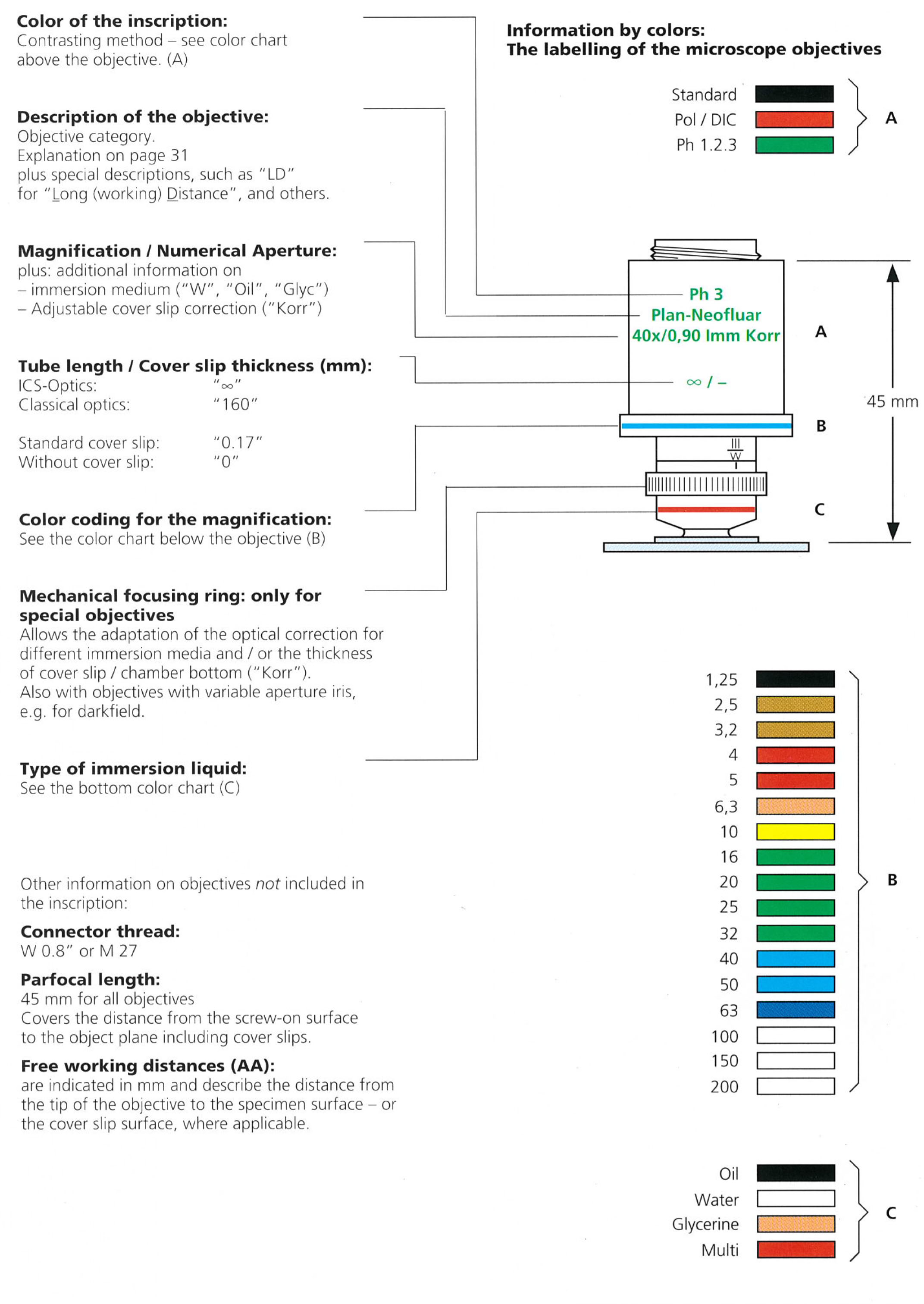
[edit] Objective Data
[edit] Refractive Index
Measurements were performed at MPI-CBG LMF, with the J457 Automatic Refractometer (RUDOPLH) for each medium, in a temperature range between 10 and 60 Celsius degrees at 1 degree steps.
[edit] Free Software


[edit] Image Acquisition
- µManager
open source software package for imaging and control of automated microscopes, refer to the µManager documentation for configurations etc.
(Windows, Mac OS, Linux)
[edit] Image Processing and Analysis
- Fiji / ImageJ
open-source software for microscopy-related tasks
- BioImageXD
open-source image analysis software
(cross platform)
- Zeiss ZEN LE
new free Zeiss software for viewing and processing *.lsm images
(Windows XP 32bit, Windows 7 32/64 bit)
- Zeiss LSM Image Browser
traditional free Zeiss software, can open database (*.mdb) and images (*.lsm) including all comments and acquisition information
(Windows only)
- Leica LAS AF Lite
free Leica software, can open *.lif and *.lei files, acquisition information are accessible
(Windows XP 32bit only)
- Olympus FV Viewer
free Olympus software, can open *.oib and *.oif files from Fluoview Confocals
(Windows XP 32bit only)
- Zeiss Axiovision LE
free version of the Axiovision software, able to open *.lsm files as well, nice batch export
(Windows only)
- IrfanView
image viewer for common file formats like *.tif and *.jpg, provides simple processing and batch conversion, slide show etc.
(Windows only)
[edit] Mailing Listserver
- The Microscopy Listserver
mailing list for all topics related to microscopy, but with a focus on electron microscopy
- Confocal List Server
Confocal Microscopy List, many subscribers and discussions, archive function
- ImageJ Mailing List
lots of discussions, good if you are interested in using ImageJ more advanced
[edit] Companies

- Carl Zeiss MicroImaging GmbH
LSM 710 and LSM 5 Live confocals, microscope frames and objectives
- Leica Microsystems
SP5(-X) and SPE confocals, microscope frames and objectives
- Olympus Microscopy
FV1000(MPE) confocal, microscope frames and objectives
- Nikon Instruments Europe B.V.
A1(R) confocal, microscope frames and objectives
- Andor Technology
Revolution, IQ software, system integration
- TILL Photonics
iMIC, Polychrome
- Improvision
Volocity
- Bitplane
Imaris
- okolab
stage incubators, cage incubators
- Ibidi
single- and multi-well dishes with cover-slip bottom, stage incubator
- Molecular Devices
MetaMorph, MetaVue
- Intelligent Imaging Innovations
FLIM, Marianas, Everest, SlideBook software, system integration
- Sutter Instrument
DG-4 lightsource, micromanipulators, accessories
- LaVision Biotec
TriMScope 2-Photon, FLIM, software, accessories
- Photometrics
cameras (CCD, EMCCD)
[edit] Our Friends and contacts
- Turku BioImaging (Finland)
- Biological imaging Development Center at UCSF
- IMP Vienna, BioOptics Facility
- McGill University Life Sciences Complex Imaging Facility
- The Leibniz Institute of Plant Genetics and Crop Plant Research (IPK) - Structural Cell Biology Group - core facility
- LMC Zürich
- Amsterdamm NL - Rob Wolthuis r.wolthuis at nki.nl