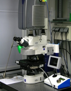
Grid LSM & WF, upr., BIOTEC
From BioDIP
(Difference between revisions)
| (14 intermediate revisions by 3 users not shown) | |||
| Line 1: | Line 1: | ||
{{Equipment | {{Equipment | ||
|system-number=BIO05 | |system-number=BIO05 | ||
| − | |system-name= | + | |system-name=Grid LSM & WF, upr., BIOTEC |
|type=Microscope | |type=Microscope | ||
|category=Real-Time Confocal | |category=Real-Time Confocal | ||
| − | |facility=Imaging@ | + | |facility=Imaging@BIOTEC/CRTD |
| − | |building= | + | |building=BIOTEC |
|room=227 | |room=227 | ||
| − | | | + | |Facility-logo=Imaging@Biotec.jpg |
| − | |description= | + | |Institute-logo=Biotec logo.png |
| + | |description=The Vt-Infinity III is dedicated for high speed confocal imaging in XYZ. The laser lines allow GFP and RFP imaging, the HBO allows common dye combinations. An EMCCD allows very fast stream acquisition of low signal. A dual-view system allows simultaneous detection of GFP and RFP in confocal mode. The use of dipping lenses makes this system especially suitable for study of embryo development. | ||
|applications=- | |applications=- | ||
|image=Bioz05.jpg | |image=Bioz05.jpg | ||
| + | |stand-name=Zeiss - Axio Imager | ||
|stand=Upright | |stand=Upright | ||
|microscope=motorized XY stage, Z-Piezo stage, fluorescence, transmitted light with manual DIC | |microscope=motorized XY stage, Z-Piezo stage, fluorescence, transmitted light with manual DIC | ||
|obj1=Zeiss EC Plan-Neofluar 10x 0.3 | |obj1=Zeiss EC Plan-Neofluar 10x 0.3 | ||
| − | |obj2=Zeiss | + | |obj2=Zeiss Achroplan 40x 0.8 W |
| − | |obj3=Zeiss | + | |obj3=Zeiss LD C-Apochromat 40x 1.1 W |
| − | + | |obj5=Zeiss Plan-Apochromat 63x 1.4 Oil | |
| − | |obj5=Zeiss | + | |obj6=Zeiss Plan-Apochromat 100x 1.4 Oil |
| − | |obj6 | + | |ill1=Fluorescence (Metal Halide, HXP, 120W) |
| − | + | ||
| − | |ill1=Fluorescence ( | + | |
|ill2=Transmitted Light (Halogen) | |ill2=Transmitted Light (Halogen) | ||
|ill3=Laser DPSS 488 nm | |ill3=Laser DPSS 488 nm | ||
|ill4=Laser DPSS 561 nm | |ill4=Laser DPSS 561 nm | ||
| − | |detection=widefield fluorescence | + | |detection=dual ccd configuration: |
| − | |reflectors=Analysator<br>GFP : EX 470/40 ; BS 495 ; EM 525/50<br>FITC : EX 480/20 ; BS 495 ; EM 535/40<br>TxRed : EX 570/20 ; BS 590 ; EM 640/40<br>DAPI (A) : EX 350/50 ; BS 400 ; EM LP 460/50 | + | Coolsnap ES for widefield fluorescence AND <br> |
| − | |features=widefield fluoerescence imaging, confocal imaging, simultaneous GFP/RFP imaging, sequential imaging, Z stacks, time-series, advanced time-series, averaging | + | Cascade II EMCCD for confocal sectioning (multi point illumination, variable pinhole size), dual channel emission beamsplitter |
| + | |reflectors=Analysator<br>GFP : EX 470/40 ; BS 495 ; EM 525/50<br>FITC : EX 480/20 ; BS 495 ; EM 535/40<br>TxRed : EX 570/20 ; BS 590 ; EM 640/40<br>DAPI (A) : EX 350/50 ; BS 400 ; EM LP 460/50 | ||
| + | |features=widefield fluoerescence imaging, confocal imaging, simultaneous GFP/RFP imaging, sequential imaging, Z stacks, time-series, advanced time-series, averaging | ||
| + | |software=MetaMorph | ||
|incubation=Stage incubator (T) | |incubation=Stage incubator (T) | ||
| − | |inv= | + | |inv=143913 |
| + | |facility_image=Imaging@Biotec.jpg | ||
| + | |logo_image=Logo_Biotec.jpg | ||
|objectives=10x/0.3<br>20x/0.5 W Ph2(Dipping)<br>40x/0.8 W (Dipping)<br>40x/3 Oil DIC<br>63x/1.3 Oil DIC<br>63x/0.95 W Ph3 (Dipping)<br>100x/1.4 Oil DIC | |objectives=10x/0.3<br>20x/0.5 W Ph2(Dipping)<br>40x/0.8 W (Dipping)<br>40x/3 Oil DIC<br>63x/1.3 Oil DIC<br>63x/0.95 W Ph3 (Dipping)<br>100x/1.4 Oil DIC | ||
|illumination=HBO lamp<br>Halogen lamp<br>491nm DPSS laser<br>561nm DPSS laser | |illumination=HBO lamp<br>Halogen lamp<br>491nm DPSS laser<br>561nm DPSS laser | ||
}} | }} | ||
Latest revision as of 11:50, 22 May 2014

|
[edit] Directions
[edit] Booking
https://techpool.biotec.tu-dresden.de/schedule/schedule.php?scheduleid=sc142df4c59eef5c
[edit] Details
| microscope | Zeiss - Axio Imager, Upright stand, motorized XY stage, Z-Piezo stage, fluorescence, transmitted light with manual DIC | ||
| objectives | |||
| illumination | |||
| detection | dual ccd configuration:
Coolsnap ES for widefield fluorescence AND | ||
| reflectors | Analysator GFP : EX 470/40 ; BS 495 ; EM 525/50 FITC : EX 480/20 ; BS 495 ; EM 535/40 TxRed : EX 570/20 ; BS 590 ; EM 640/40 DAPI (A) : EX 350/50 ; BS 400 ; EM LP 460/50 | ||
| features | widefield fluoerescence imaging, confocal imaging, simultaneous GFP/RFP imaging, sequential imaging, Z stacks, time-series, advanced time-series, averaging | ||
| software | MetaMorph | ||
| incubation | Stage incubator (T) | ||
| links |
| ||
| inv.nr. | 143913 |
