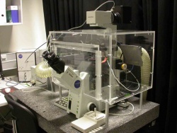
L5 - Olympus ScanR+CellR
From BioDIP
(Difference between revisions)
| Line 1: | Line 1: | ||
{{Equipment | {{Equipment | ||
| − | | system-number | + | |system-number=MPI24 |
| − | | system-name | + | |system-name=Olympus ScanR + CellR |
| − | | category | + | |type=Microscope |
| − | + | |category=Wide-Field | |
| − | + | |facility=MPI-CBG LMF | |
| − | | facility | + | |building=MPI-CBG |
| − | + | |room=U50 | |
| − | | building | + | |facility_image=logo-lmf.jpg |
| − | | room | + | |description=Motorized inverted wide-field system with temperature and CO2 cage incubator. |
| − | | description | + | |applications=long-term observation of living cells, Fast multi-fluorescence 3-D time-lapse, Fast and precise image acquisition and analysis, robust automated object detection and cell separation, visual correlation of cell images and analysis results. |
| − | | applications | + | |image=MPI24 .jpg |
| − | | microscope | + | |stand=Inverted |
| − | |obj1= | + | |microscope=motorized XY stage, motorized Z-drive, fluorescence, transmitted light |
| − | |obj2= | + | |obj1=- |
| − | |obj3= | + | |obj2=- |
| − | |obj4= | + | |obj3=- |
| − | |obj5= | + | |obj4=- |
| − | |obj6= | + | |obj5=- |
| + | |obj6=- | ||
|obj7=- | |obj7=- | ||
|obj8=- | |obj8=- | ||
| − | |ill1=Halogen | + | |ill1=Transmitted Light (Halogen) |
| − | |ill2= | + | |ill2=Fluorescence (Xenon) |
|ill3=- | |ill3=- | ||
|ill4=- | |ill4=- | ||
| Line 28: | Line 29: | ||
|ill7=- | |ill7=- | ||
|ill8=- | |ill8=- | ||
| − | | detection | + | |detection=Hamamatsu Camera ORCA ER, progressive scan interline CCD, 1344 x 1024 pixels, 6.45 um pixel size, 14.75 MHz digitization, 8 e- rms readout noise, 8 fps @ full frame, 18.000 e- full well capacity, 2250:1 dynamic range, peltier cooling (-20 degrees C @ 20 degrees C ambient), 12 bit ADC, 0.1 electron/pixel/sec dark current, up to 10x analog gain, offset functions |
| − | | reflectors | + | |reflectors=MT20 Excitation Unit<br> - EXCITATION FILTERS <br> 403/ 492 / 572 / 387/ 485 / 560/ 650 <br> |
- DICHROICS AND EMISSION FILTERS <br> * POSITION 1: <br> D 405-435/450-485/510-550/590-675 <br>X 450-475/515-545/600-665 <br> * POSITION 2:<BR> D QBP 410-465, 504-538, 581-632, 669-743 <BR> M QBP 415-461, 507-533, 586-625, 673-722 <BR> * POSITION 3: EMPTY <BR> * POSITION 4: EMPTY<BR> * POSITION 5:<BR> D 485-570/605-690<br> X500-560/615-675<br> * POSITION 6<BR> D 440-490/510-575/610-675 <br>X 450-480/525-560/615-660 | - DICHROICS AND EMISSION FILTERS <br> * POSITION 1: <br> D 405-435/450-485/510-550/590-675 <br>X 450-475/515-545/600-665 <br> * POSITION 2:<BR> D QBP 410-465, 504-538, 581-632, 669-743 <BR> M QBP 415-461, 507-533, 586-625, 673-722 <BR> * POSITION 3: EMPTY <BR> * POSITION 4: EMPTY<BR> * POSITION 5:<BR> D 485-570/605-690<br> X500-560/615-675<br> * POSITION 6<BR> D 440-490/510-575/610-675 <br>X 450-480/525-560/615-660 | ||
| − | | features | + | |features=Dual System. It can work as Scan R or as CELL R <br>SCAN R <br> Modular microscope-based imaging platform designed for fully automated image acquisition and data analysis of biological samples. scan^R can handle many different formats e.g. multi-well plates, slides or custom-built arrays. The unmatched flexibility and open design make it equally adept at routine and advanced applications. With its powerful analysis module for biological functional assays, it is the perfect tool for assay development and high-content screening. This provides complex image analysis and advanced data evaluation. <br> CELL R<br> consists of an all-in-one illumination system MT20, highly sensitive digital cameras and a hyper-precision hardware control board. |
| − | + | |incubation=cage incubator (T+CO2) | |
| − | | incubation | + | |link1=- |
| − | | link1 | + | |link2=- |
| − | | link2 | + | |inv=405679 |
| − | | inv | + | |objectives=# UPlanSApo 4X N.A 0.16 |
| + | # UPlanSApo 10X N.A 0.4 | ||
| + | # UPlanFluor 20X N.A 0.5 | ||
| + | # UPlanSApo 40X N.A 0.9 | ||
| + | # UPlanSApo 60X N.A 1.35 Oil | ||
| + | # UPlanFluor 60x N.A. 1.25 Oil Ph3 Iris (loan!!) | ||
| + | |illumination=- | ||
}} | }} | ||
Revision as of 16:29, 11 September 2009
Directions
[[image:|150px|text-bottom|right]]
Booking
https://python-srv1.mpi-cbg.de/lmf-ipf/cgi-bin/index.py
Details
| microscope | , Inverted stand, motorized XY stage, motorized Z-drive, fluorescence, transmitted light | ||
| objectives | |||
| illumination | |||
| detection | Hamamatsu Camera ORCA ER, progressive scan interline CCD, 1344 x 1024 pixels, 6.45 um pixel size, 14.75 MHz digitization, 8 e- rms readout noise, 8 fps @ full frame, 18.000 e- full well capacity, 2250:1 dynamic range, peltier cooling (-20 degrees C @ 20 degrees C ambient), 12 bit ADC, 0.1 electron/pixel/sec dark current, up to 10x analog gain, offset functions | ||
| reflectors | MT20 Excitation Unit - EXCITATION FILTERS 403/ 492 / 572 / 387/ 485 / 560/ 650 - DICHROICS AND EMISSION FILTERS | ||
| features | Dual System. It can work as Scan R or as CELL R SCAN R Modular microscope-based imaging platform designed for fully automated image acquisition and data analysis of biological samples. scan^R can handle many different formats e.g. multi-well plates, slides or custom-built arrays. The unmatched flexibility and open design make it equally adept at routine and advanced applications. With its powerful analysis module for biological functional assays, it is the perfect tool for assay development and high-content screening. This provides complex image analysis and advanced data evaluation. CELL R consists of an all-in-one illumination system MT20, highly sensitive digital cameras and a hyper-precision hardware control board."Dual System. It can work as Scan R or as CELL R <br />SCAN R <br /> Modular microscope-based imaging platform designed for fully automated image acquisition and data analysis of biological samples. scan^R can handle many different formats e.g. multi-well plates, slides or custom-built arrays. The unmatched flexibility and open design make it equally adept at routine and advanced applications. With its powerful analysis module for biological functional assays, it is the perfect tool for assay development and high-content screening. This provides complex image analysis and advanced data evaluation. <br /> CELL R<br /> consists of an all-in-one illumination system MT20, highly sensitive digital cameras and a hyper-precision hardware control board." cannot be used as a page name in this wiki. | ||
| software | |||
| incubation | cage incubator (T+CO2) | ||
| links |
| ||
| inv.nr. | 405679 |
