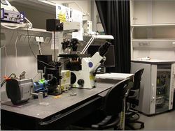
SD1 - Andor Spinning Disc
From BioDIP
(Difference between revisions)
(edit after major upgrade: scan head, objectives, iQ version) |
|||
| Line 7: | Line 7: | ||
|building=MPI-CBG | |building=MPI-CBG | ||
|room=038B | |room=038B | ||
| − | |description=Fast, "real-time" confocal imaging. Laser equipment: 488 nm solid-state Coherent Sapphire (75mW) | + | |Facility-logo=MPI-CBG LMF.jpg |
| − | |applications=confocal imaging of EGFP | + | |description=Fast, "real-time" confocal imaging. Laser equipment: 488 nm solid-state Coherent Sapphire (75mW), 561 nm solid-state Cobolt Jive (75mW) + 640 Coherent Cube (40mW) . |
| + | |applications=confocal imaging of EGFP, mCherry, dTomato, Draq5 and the like with high temporal and spatial resolution. | ||
|image=MPI19.jpg | |image=MPI19.jpg | ||
|stand-name=Olympus - IX71 | |stand-name=Olympus - IX71 | ||
| Line 14: | Line 15: | ||
|microscope=manual, 1.6x OptoVar, fast piezo objective z-positioner (Physik Instrumente - PI), Prior ProScanIII xy scanning stage | |microscope=manual, 1.6x OptoVar, fast piezo objective z-positioner (Physik Instrumente - PI), Prior ProScanIII xy scanning stage | ||
|obj1=Olympus UPlanSApo 10x 0.4 | |obj1=Olympus UPlanSApo 10x 0.4 | ||
| − | |obj2=Olympus UPlanSApo 60x 1.20 W | + | |obj2=Olympus UPlanApo 20x 0.7 |
| − | | | + | |obj3=Olympus UPlan FluarN 60x 0.9 |
| − | + | |obj4=Olympus UPlanSApo 60x 1.20 W | |
| + | |obj5=Olympus UPlanSApo 100x 1.4 Oil | ||
|ill1=Transmitted Light (Halogen) | |ill1=Transmitted Light (Halogen) | ||
|ill2=Fluorescence (HBO) | |ill2=Fluorescence (HBO) | ||
|ill3=Laser DPSS 488 nm | |ill3=Laser DPSS 488 nm | ||
|ill4=Laser DPSS 561 nm | |ill4=Laser DPSS 561 nm | ||
| − | |detection=Spinning disc scan head Yokogawa | + | |ill5=Laser DPSS 640 nm |
| − | |reflectors=Optical Insight DualView image splitter: <br>holder for GFP/mCherry: BL 525/40; BS 565; ET 605/70<br>holder for GFP/RFP: HC 520/35; BS 565; HC 628/40<br>Sutter filter wheel: <br>position 0: 624/40, 1: open, 2: 525/30, 3: razor edge 568LP, 4: dual emitter: 512/23+630/91, 5: razor edge 488LP, 6: old CSU dual emitter: 525/30 + 650/?, 7: 605/70, 8: 445/40, 9: 716/40<br> microscope filter turret:<br> position 1: GFP cube, 2: RFP cube, 3: dual GFP + RFP cube, 4: DAPI cube, 5: empty, 6: DIC analyzer | + | |detection=Spinning disc scan head Yokogawa CSU-X1: dichromatic mirrors: [http://www.semrock.com/FilterDetails.aspx?id=Di01-T405/488/568/647-13x15x0.5 T-405/488/568/647]<br> [http://www.semrock.com/FilterDetails.aspx?id=Di01-T405/488/561-13x15x0.5 T-405/488/561] <br>physical pinhole radius: 24um, physical pinhole spacing: 240um; back-projected pinhole radius: 0.15um, back-projected pinhole spacing: 1.5um (with 100x objective, 1.6x optovar)<br>Andor iXon EM+ DU-897 BV back illuminated EMCCD; pixel size of EMCCD chip: 16um<br>image pixel size (measured again after major scope repair on March 7th 2011): with 100x objective: 0.175um (was 0.178um before 20110307); 100x obj. and 1.6x optovar: 0.109um (was 0.111um before 20110307); 60x obj.: 0.289um (was 0.288um before); 60x obj. and 1.6x opt.: 0.180um (was 0.181um before 20110307); 10x obj. : 1.747um, 10x obj. and 1.6x opt.: 1.089um<br>camera projecting lens has about 0.92x (was 0.9x before 2011_03_07) magnification calculated from the measured pixel sizes |
| + | |reflectors=Optical Insight DualView image splitter (currently not installed): <br>holder for GFP/mCherry: BL 525/40; BS 565; ET 605/70<br>holder for GFP/RFP: HC 520/35; BS 565; HC 628/40<br>Sutter filter wheel: <br>position 0: 624/40, 1: open, 2: 525/30, 3: razor edge 568LP, 4: dual emitter: 512/23+630/91, 5: razor edge 488LP, 6: old CSU dual emitter: 525/30 + 650/?, 7: 605/70, 8: 445/40, 9: 716/40<br> microscope filter turret:<br> position 1: GFP cube, 2: RFP cube, 3: dual GFP + RFP cube, 4: DAPI cube, 5: empty, 6: DIC analyzer | ||
|features=- | |features=- | ||
| − | |software=iQ | + | |software=iQ 2.5.1 |
|incubation=Stage incubator (T) + objective heater | |incubation=Stage incubator (T) + objective heater | ||
|link0=https://ifn.mpi-cbg.de/wiki/ifn/index.php/MPI-SD1_-_Olympus_Andor_Spinning_Disc_-_startup_%26_shutdown | |link0=https://ifn.mpi-cbg.de/wiki/ifn/index.php/MPI-SD1_-_Olympus_Andor_Spinning_Disc_-_startup_%26_shutdown | ||
Revision as of 16:13, 5 April 2012

|
Directions
[[image:|150px|text-bottom|right]]
Booking
https://python-srv1.mpi-cbg.de/lmf-ipf/cgi-bin/index.py
Details
| microscope | Olympus - IX71, Inverted stand, manual, 1.6x OptoVar, fast piezo objective z-positioner (Physik Instrumente - PI), Prior ProScanIII xy scanning stage | ||
| objectives | |||
| illumination | |||
| detection | [[detection::Spinning disc scan head Yokogawa CSU-X1: dichromatic mirrors: T-405/488/568/647 T-405/488/561 physical pinhole radius: 24um, physical pinhole spacing: 240um; back-projected pinhole radius: 0.15um, back-projected pinhole spacing: 1.5um (with 100x objective, 1.6x optovar) Andor iXon EM+ DU-897 BV back illuminated EMCCD; pixel size of EMCCD chip: 16um image pixel size (measured again after major scope repair on March 7th 2011): with 100x objective: 0.175um (was 0.178um before 20110307); 100x obj. and 1.6x optovar: 0.109um (was 0.111um before 20110307); 60x obj.: 0.289um (was 0.288um before); 60x obj. and 1.6x opt.: 0.180um (was 0.181um before 20110307); 10x obj. : 1.747um, 10x obj. and 1.6x opt.: 1.089um camera projecting lens has about 0.92x (was 0.9x before 2011_03_07) magnification calculated from the measured pixel sizes]] | ||
| reflectors | Optical Insight DualView image splitter (currently not installed): holder for GFP/mCherry: BL 525/40; BS 565; ET 605/70 holder for GFP/RFP: HC 520/35; BS 565; HC 628/40 Sutter filter wheel: position 0: 624/40, 1: open, 2: 525/30, 3: razor edge 568LP, 4: dual emitter: 512/23+630/91, 5: razor edge 488LP, 6: old CSU dual emitter: 525/30 + 650/?, 7: 605/70, 8: 445/40, 9: 716/40 microscope filter turret: position 1: GFP cube, 2: RFP cube, 3: dual GFP + RFP cube, 4: DAPI cube, 5: empty, 6: DIC analyzer | ||
| features | - | ||
| software | iQ 2.5.1 | ||
| incubation | Stage incubator (T) + objective heater | ||
| links |
| ||
| inv.nr. | 0 10.1.65.208 |
