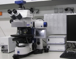
Zeiss ApoTome - DZNE W1
From BioDIP
(Difference between revisions)
| Line 10: | Line 10: | ||
|Institute-logo=Logo-DZNE.jpg | |Institute-logo=Logo-DZNE.jpg | ||
|description=It is designed for routine work on fixed samples. Optical sectioning can be obtained on fixed samples by the use of the Apotome. | |description=It is designed for routine work on fixed samples. Optical sectioning can be obtained on fixed samples by the use of the Apotome. | ||
| − | |applications= | + | |applications=fixed samples, fluorescently labeled samples (structured illumination possible), histologically fixed samples (no strucutred illumination), big samples |
| − | + | ||
| − | + | ||
| − | + | ||
| − | + | ||
|image=Zeiss ApoTome - DZNE 01.jpg | |image=Zeiss ApoTome - DZNE 01.jpg | ||
|stand-name=Zeiss - Axio Imager.M2 | |stand-name=Zeiss - Axio Imager.M2 | ||
| Line 35: | Line 31: | ||
<br> | <br> | ||
*FS Analysator for DIC and Transmission | *FS Analysator for DIC and Transmission | ||
| − | |features= | + | |features=sequential imaging, Brightfield and Fluorescence overlay, timelapse, z-stack, tile scans, multi position experiments, complex experiments via Experiment Designer, optical sectioning |
| − | + | ||
| − | + | ||
| − | + | ||
| − | + | ||
| − | + | ||
| − | + | ||
| − | + | ||
| − | + | ||
|software=ZEN Blue 2012 | |software=ZEN Blue 2012 | ||
|incubation=Not available | |incubation=Not available | ||
|inv=0 | |inv=0 | ||
}} | }} | ||
Latest revision as of 10:00, 3 August 2016

|
[edit] Directions
[edit] Booking
https://techpool.biotec.tu-dresden.de/schedule/schedule.php?scheduleid=sc14ffd3aa3e44c3
[edit] Details
| microscope | Zeiss - Axio Imager.M2, Upright stand, motorized XY stage (Märzhäuser SMC2009), motorized z-drive, fluorescence, transmitted light with DIC, Darkfield b/w epifluorescence acquisition, Optical Sectioning (ApoTome2) | ||
| objectives | |||
| illumination | |||
| detection |
| ||
| reflectors |
| ||
| features | sequential imaging, Brightfield and Fluorescence overlay, timelapse, z-stack, tile scans, multi position experiments, complex experiments via Experiment Designer, optical sectioning | ||
| software | ZEN Blue 2012 | ||
| incubation | Not available | ||
| links |
| ||
| inv.nr. | 0 |

