For imaging your histologically stained samples the system now also features a coloured camera."It is designed for routine work on fixed samples. Optical sectioning can be obtained on fixed samples by the use of the Apotome.
For imaging your histologically stained samples the system now also features a coloured camera." cannot be used as a page name in this wiki.
Suitable for
- fixed samples
- fluorescently labeled samples (structured illumination possible)
- histologically fixed samples (no strucutred illumination)"<br />
- fixed samples
- fluorescently labeled samples (structured illumination possible)
- histologically fixed samples (no strucutred illumination)" cannot be used as a page name in this wiki..
|
Directions
Booking
https://techpool.biotec.tu-dresden.de/schedule/schedule.php?scheduleid=sc14ffd3aa3e44c3
Details
| microscope
|
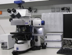
Zeiss - Axio Imager.M2, Upright stand, motorized XY stage (Märzhäuser SMC2009), motorized z-drive, fluorescence, transmitted light with DIC, Darkfield b/w epifluorescence acquisition, Optical Sectioning (ApoTome2)
|
| objectives
|
- Zeiss EC Plan Neofluar 5x 0.16
- Zeiss Plan-Apochromat 10x 0.45
- Zeiss Plan-Apochromat 20x 0.8
- Zeiss Plan-Apochromat 40x 0,95 korr.
- Zeiss EC Plan-Neofluar 100x 1.3 Oil
-
-
-
-
-
|
| illumination
|
- Fluorescence (Metal Halide, HXP, 120W)
- Transmitted Light (Halogen)
-
-
-
-
-
-
-
-
|
| detection
|
- Axiocam MRm rev.3 (1388*1040, 6,45*6,45µm)
|
| reflectors
|
- FS 49 (DAPI) : EX BP G365; BS 395; EM BP 445/50
- FS 38 HE (GFP) : EX BP 470/40; BS 495; EM BP 525/50
- FS 20 (Rhodamine) : EX BP 546/12; BS 560; BP 575-640
- FS 50 (Cy5) : EX BP 640/30 ; BS 660 ; EM 690/50
- FS Analysator for DIC and Transmission
|
| features
|
- sequential imaging
- Brightfield and Fluorescence overlay
- timelapse
- z-stack
- tile scans
- multi position experiments
- complex experiment via Experiment Designer
- optical sectioning
- histollogical imaging"<br />
- sequential imaging
- Brightfield and Fluorescence overlay
- timelapse
- z-stack
- tile scans
- multi position experiments
- complex experiment via Experiment Designer
- optical sectioning
- histollogical imaging" cannot be used as a page name in this wiki.
|
| software
|
ZEN Blue 2012
|
| incubation
|
Not available
|
| links
|
- [ manual]
- [ laser power values]
|
- [ beam path]
- [ advanced system check]
|
|
| inv.nr.
|
0
|

