ASC Fluorescence and Brightfield Imaging
From BioDIP
(Difference between revisions)
(→images) |
|||
| Line 12: | Line 12: | ||
== images == | == images == | ||
<gallery> | <gallery> | ||
| − | File:Conva10xok.jpg with bar.jpg|Convallaria, 10x/0.45 objective, 488 nm excitation, BP505-550 detection | + | File:Conva10xok.jpg with bar.jpg|Convallaria, Zeiss LSM 510, 10x/0.45 objective, 488 nm excitation, BP505-550 detection, pinhole: 1 airy unit |
| − | File:Conva10xok-1.jpg with bar.jpg|Convallaria, 10x/0.45 objective, 488 nm illumination, transmitted light detection | + | File:Conva10xok-1.jpg with bar.jpg|Convallaria, Zeiss LSM 510, 10x/0.45 objective, 488 nm illumination, transmitted light detection |
</gallery> | </gallery> | ||
__NOTOC__ | __NOTOC__ | ||
Revision as of 14:54, 30 September 2009
First step of the system check. The purpose is to test the general performance of the system: Do the basic functions work fine? Does the acquisition software behave as it should? Can one set up Koehler illumination? Are the transmitted and the reflected beam-paths ok? Although this test does not represent high-resolution work, it might reveal general problems with the system.
requirements
- basic sample with fluorescence and contrast in transmitted light (i.e. Zeiss/Leica Convallaria demo sample)
acquisition
- set up the beam-path for fluorescence and transmitted light
- use basic air objective (i.e. 10x/0.4)
- acquire two images in basic resolution (i.e. 512x512)
analysis
- Does the illumination look reasonably homogeneous over the whole field of view in both images?
- Is the transmitted light image free of dirt particles?
- Are both images free of artifacts?
images
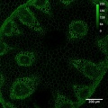
Convallaria, Zeiss LSM 510, 10x/0.45 objective, 488 nm excitation, BP505-550 detection, pinhole: 1 airy unit
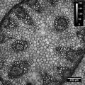
Convallaria, Zeiss LSM 510, 10x/0.45 objective, 488 nm illumination, transmitted light detection

