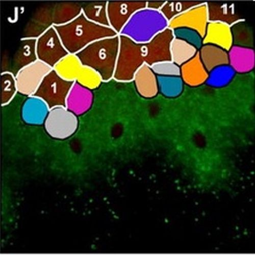
Origin and shaping of the laterality organ in zebrafish.
Handedness of the vertebrate body plan critically depends on transient embryonic structures/organs that generate cilia-dependent leftward fluid flow within constrained extracellular environments. Although the function of ciliated organs in laterality determination has been extensively studied, how they are formed during embryogenesis is still poorly understood. Here we show that Kupffer's vesicle (KV), the zebrafish organ of laterality, arises from a surface epithelium previously thought to adopt exclusively extra-embryonic fates. Live multi-photon confocal imaging reveals that surface epithelial cells undergo Nodal/TGFbeta signalling-dependent ingression at the dorsal germ ring margin prior to gastrulation, to give rise to dorsal forerunner cells (DFCs), the precursors of KV. DFCs then migrate attached to the overlying surface epithelium and rearrange into rosette-like epithelial structures at the end of gastrulation. During early somitogenesis, these epithelial rosettes coalesce into a single rosette that differentiates into the KV with a ciliated lumen at its apical centre. Our results provide novel insights into the morphogenetic transformations that shape the laterality organ in zebrafish and suggest a conserved progenitor role of the surface epithelium during laterality organ formation in vertebrates.

- Development 2008 Aug 17;135(16):2807-13
- 2008
- Developmental Biology
- 18635607
- PubMed
Enabled by:
