Ancestral vascular lumen formation via basal cell surfaces.
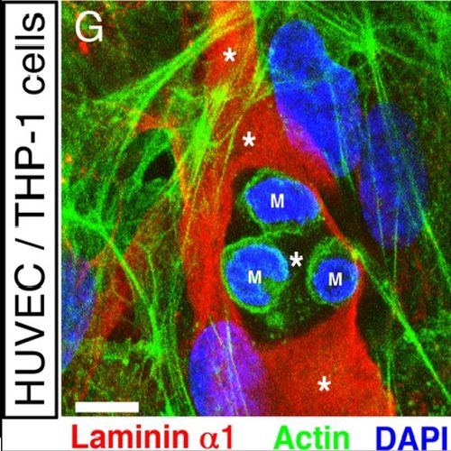
The cardiovascular system of bilaterians developed from a common ancestor. However, no endothelial cells exist in invertebrates demonstrating that primitive cardiovascular tubes do not require this vertebrate-specific cell type in order to form. This raises the question of how cardiovascular tubes form in invertebrates? Here we discovered that in the invertebrate cephalochordate amphioxus, the basement membranes of endoderm and mesoderm line the lumen of the major vessels, namely aorta and heart. During amphioxus development a laminin-containing extracellular matrix (ECM) was found to fill the space between the basal cell surfaces of endoderm and mesoderm along their anterior-posterior (A-P) axes. Blood cells appear in this ECM-filled tubular space, coincident with the development of a vascular lumen. To get insight into the underlying cellular mechanism, we induced vessels in vitro with a cell polarity similar to the vessels of amphioxus. We show that basal cell surfaces can form a vascular lumen filled with ECM, and that phagocytotic blood cells can clear this luminal ECM to generate a patent vascular lumen. Therefore, our experiments suggest a mechanism of blood vessel formation via basal cell surfaces in amphioxus and possibly in other invertebrates that do not have any endothelial cells. In addition, a comparison between amphioxus and mouse shows that endothelial cells physically separate the basement membranes from the vascular lumen, suggesting that endothelial cells create cardiovascular tubes with a cell polarity of epithelial tubes in vertebrates and mammals.

- PLoS ONE 2009 Jan 06;4(1):e4132
- 2009
- Developmental Biology
- 19125185
- PubMed
Enabled by:
