Long-bone critical-size defects treated with tissue-engineered polycaprolactone-co-lactide scaffolds: a pilot study on rats.
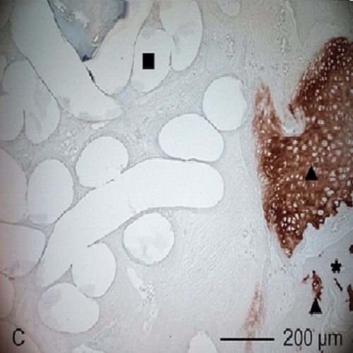
The aim of this study was to evaluate the osteogenic potential of embroidered, tissue-engineered polycaprolactone-co-lactide (trade name: PCL) scaffolds for the reconstruction of large bone defects. Ten piled-up PCL scaffolds were implanted in femura with a critical size defect of immunodeficient nude rats for 12 weeks [n = 4, group 1: noncoated, group 2: collagen I (coll I), group 3: collagen I/chondroitin sulfate (coll I/CS), and group 4: collagen I/chondroitin sulfate/human mesenchymal stem cells (coll I/CS/hMSC)]. X-ray examination, computer tomography, and histological analyses of the explanted scaffold pads were performed. The quantification of the bone volume ratio showed a significantly higher rate of new bone formation at coll I/CS-coated scaffolds compared with the other groups. Histological investigations revealed that the defect reconstruction started from the peripheral bone ends and incorporated into the scaffold material. Additionally seeded hMSC on coll I/CS-coated scaffolds showed a higher matrix deposition inside the implant but no higher bone formation was observed. These data imply that the coll I/CS-coated PCL scaffolds have the highest potential for treating critical size defects. The scaffolds, being variable in size and structure, can be adapted to any bone defect.

- J Biomed Mater Res A 2010 Dec 1;95(3):964-72
- 2010
- Medical Biology
- 20824650
- PubMed
Enabled by:
