Wnt interaction and extracellular release of prominin-1/CD133 in human malignant melanoma cells.
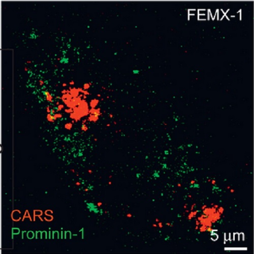
Prominin-1 (CD133) is the first identified gene of a novel class of pentaspan membrane glycoproteins. It is expressed by various epithelial and non-epithelial cells, and notably by stem and cancer stem cells. In non-cancerous cells such as neuro-epithelial and hematopoietic stem cells, prominin-1 is selectively concentrated in plasma membrane protrusions, and released into the extracellular milieu in association with small vesicles. Previously, we demonstrated that prominin-1 contributes to melanoma cells pro-metastatic properties and suggested that it may constitute a molecular target to prevent prominin-1-expressing melanomas from colonizing and growing in lymph nodes and distant organs. Here, we report that three distinct pools of prominin-1 co-exist in cultures of human FEMX-I metastatic melanoma. Morphologically, in addition to the plasma membrane localization, prominin-1 is found within the intracellular compartments, (e.g., Golgi apparatus) and in association with extracellular membrane vesicles. The latter prominin-1-positive structures appeared in three sizes (small, ?40 nm; intermediates ~40-80 nm, and large, >80 nm). Functionally, the down-regulation of prominin-1 in FEMX-I cells resulted in a significant reduction of number of lipid droplets as observed by coherent anti-Stokes Raman scattering image analysis and Oil red O staining, and surprisingly in a decrease in the nuclear localization of beta-catenin, a surrogate marker of Wnt activation. Moreover, the T-cell factor/lymphoid enhancer factor (TCF/LEF) promoter activity was 2 to 4 times higher in parental than in prominin-1-knockdown cells. Collectively, our results point to Wnt signaling and/or release of prominin-1-containing membrane vesicles as mediators of the pro-metastatic activity of prominin-1 in FEMX-I melanoma.

- Exp. Cell Res. 2013 Apr 1;319(6):810-9
- 2013
- Cell Biology
- 23318676
- PubMed
Enabled by:
