Gallery
Here all images used on this website can be found including the author, the title and a short description. If you want to know more details about any of those, just get in touch with us!
Light Microscopy Images
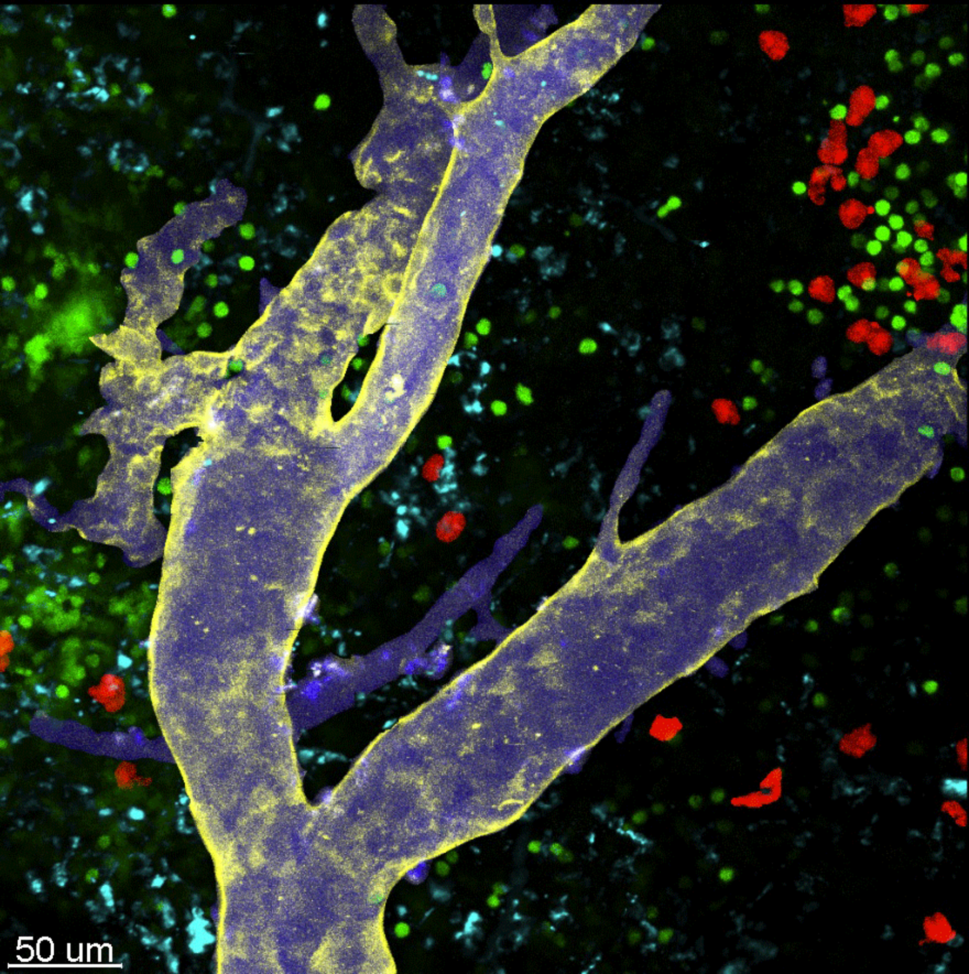
 supported by LMF CMCB") © Josh Currie - CRTD
© Josh Currie - CRTD
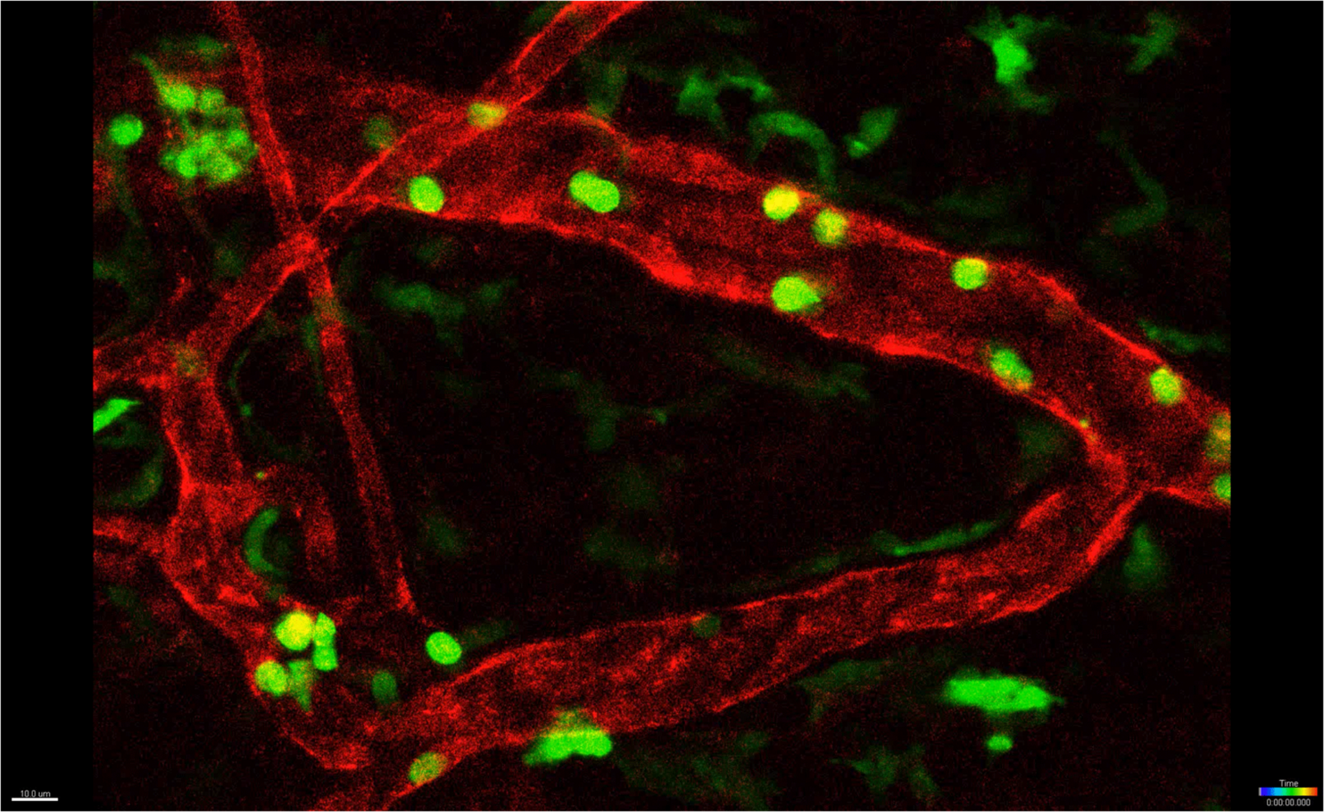
 © Judith Konantz - CRTD
 marked with GFP. Note that the NE does not break down during cell division.") © Isabel Raabe - MPI-CBG
© Isabel Raabe - MPI-CBG

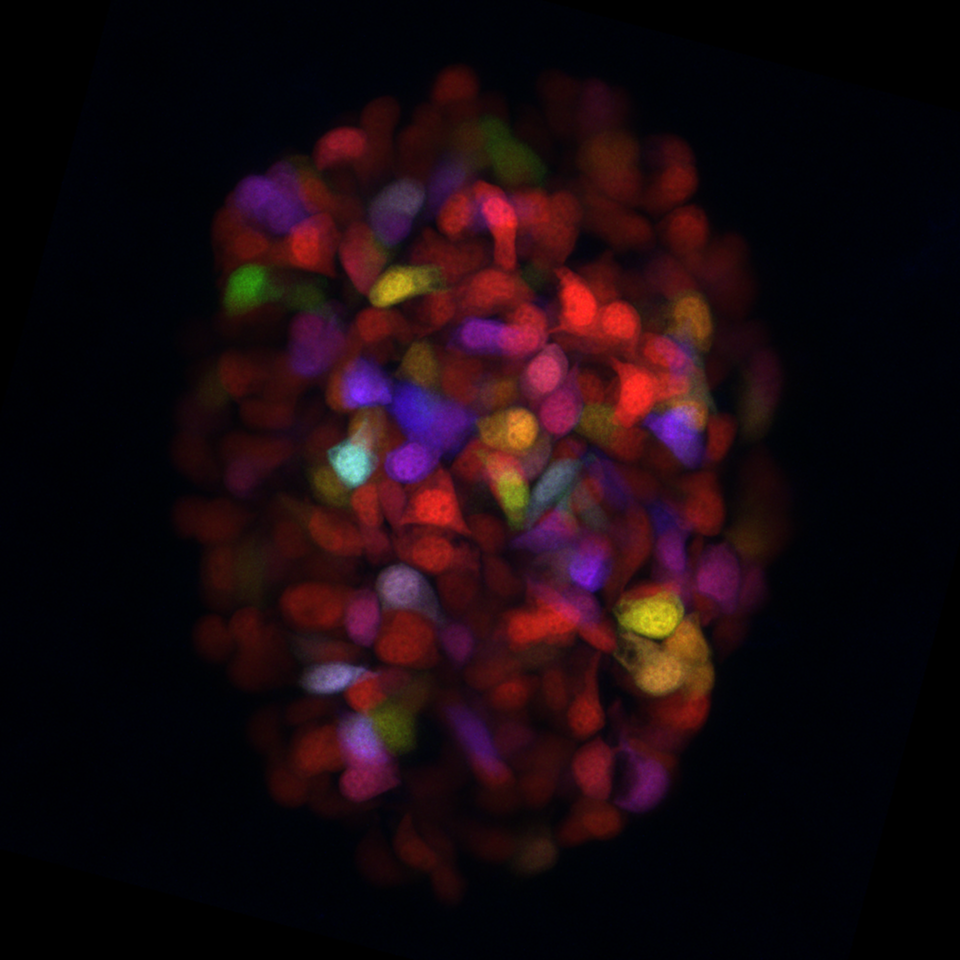
 © Kathleen Roensch - CRTD

 © Edgar Büttner - BIOTEC
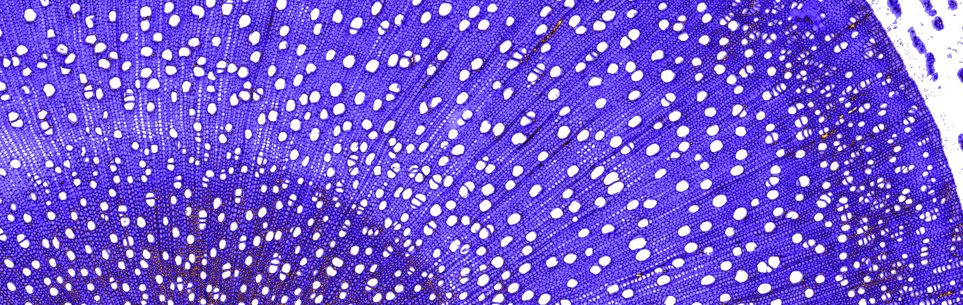


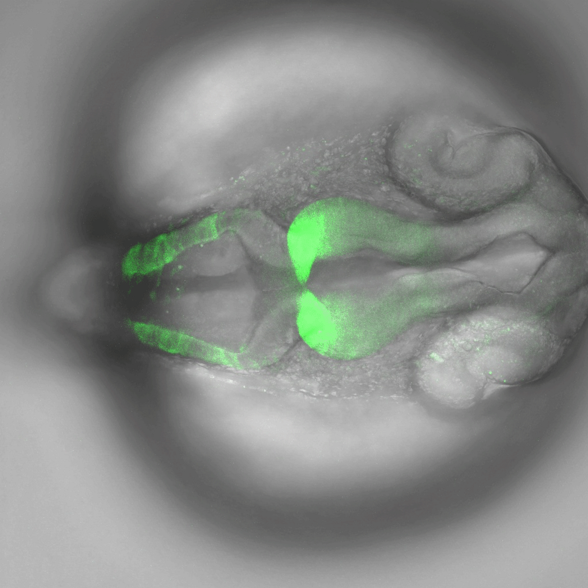
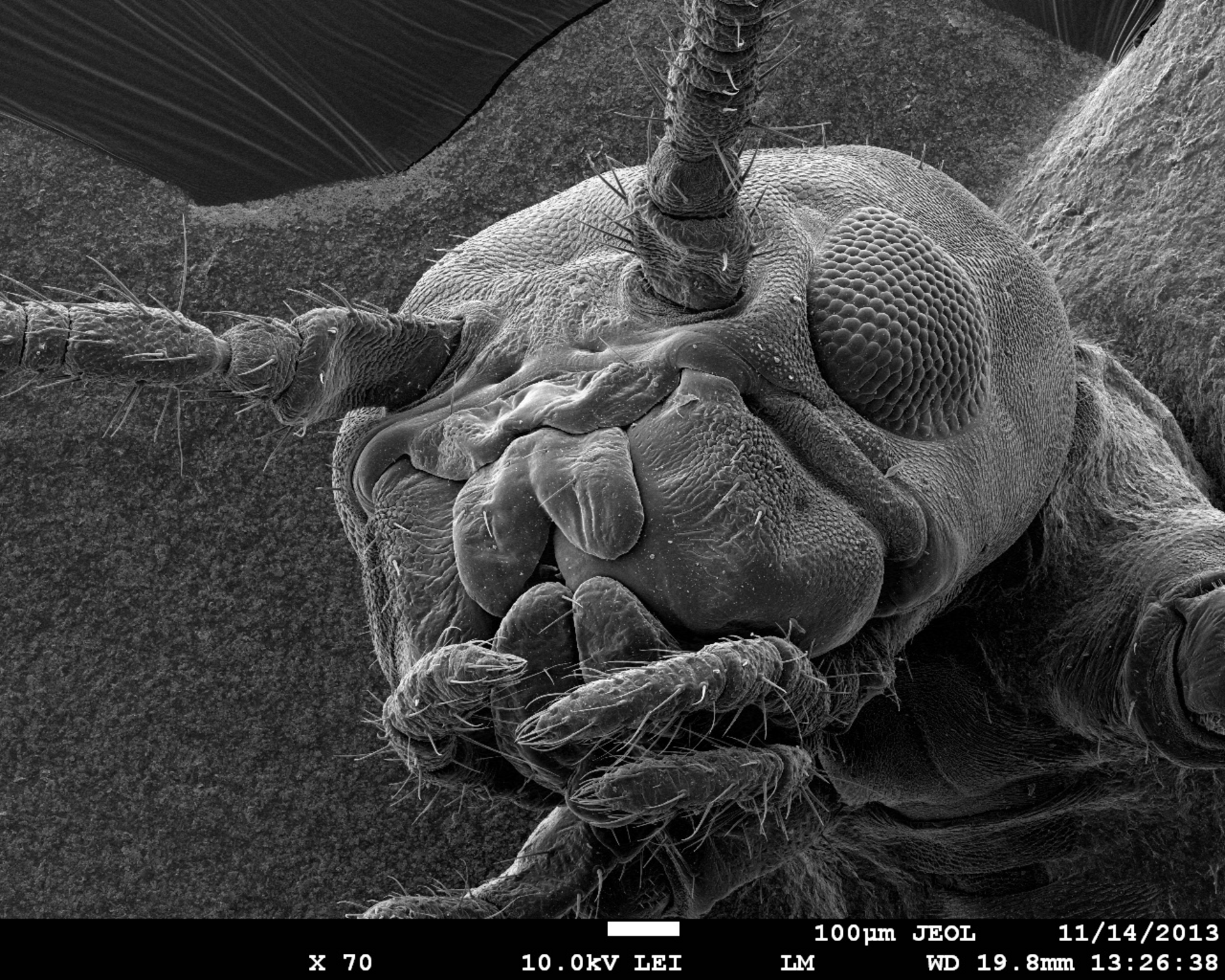
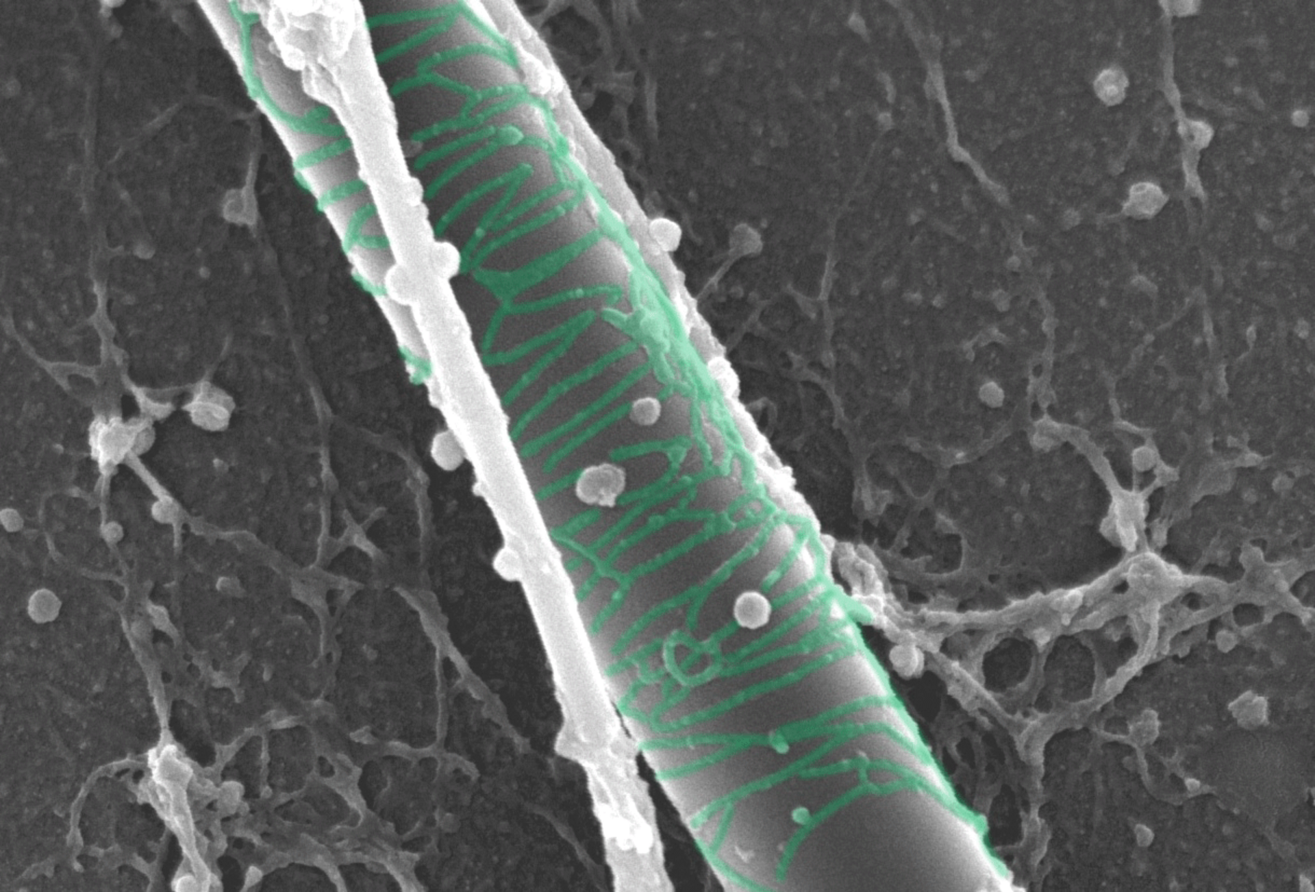
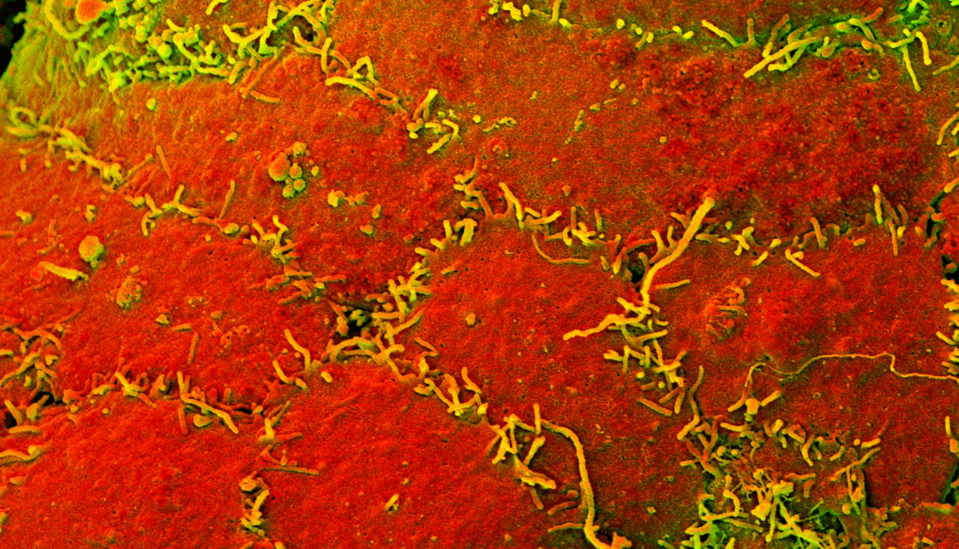
Image Processing
 © Steffen Vogler & Fanny Boehme - DZNE